
|
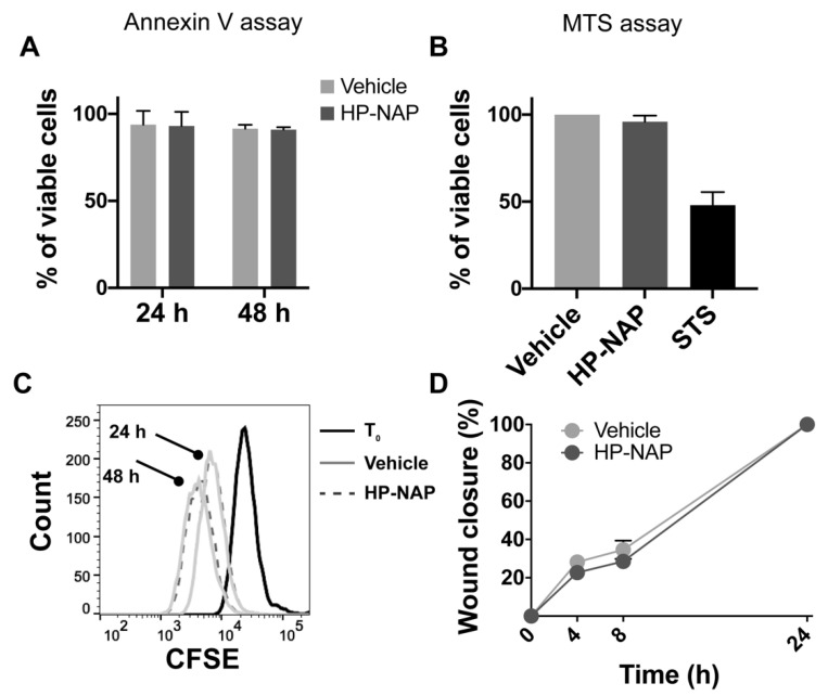
Figure 3
HP-NAP has no direct effect on melanoma cells. (A) M121224 cells were treated with HP-NAP or saline (vehicle) and after 24 and 48 h, apoptosis was evaluated by annexin V staining. (B) M121224 cells were treated with HP-NAP, saline (vehicle) or staurosporine (STS) as positive control; cytotoxicity was evaluated by MTS assay. Values are reported as percentage of viable cells ± SEM of three independent experiments. (C) M121224 cells were labeled with CSFE and treated with HP-NAP or saline (vehicle) for 24 and 48 h. Cells were harvested, washed, resuspended in saline and analyzed by flow cytometry. Representative histograms from two independent experiments are reported. (D) Cell migration was evaluated after a 24 h incubation with HP-NAP in serum-free RPMI (supplemented with 0.1% BSA). Cells incubated in medium with the same volume of saline were considered as control (vehicle). 0.1 × 10⁶ M121224 cells were seeded in a 24-well culture dish. Scratches were created once cells reached confluence. At time zero and after 4, 8 and 24 h, wound closures were photographed under a microscope. Migration rate was expressed as percentage of wound closure (0%: T0 after wound; 100%: completely repaired).