
|
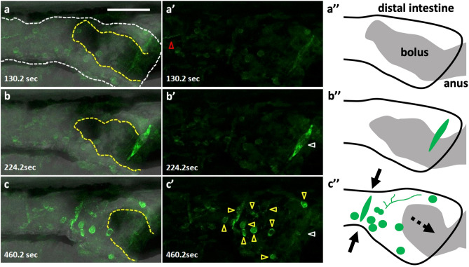
Figure 3
Peristaltic reflex visualized by Ca2+ imaging of a variety of cell types including putative enteric neurons and circular smooth muscles. Live images of putative enteric neurons and circular smooth muscles expressing GCaMP3 at 8 dpf in Tg(hsp70: Gal4); Tg(UAS: GCaMP3), heat-shocked at 6 dpf. The passage of the bolus was blocked at the anus by agarose gel, in an artificial condition of constipation. (a–a’’) the phase when most of the activity is arrested but an activated fiber with a distinct rhythm extends from the oral side (red triangle). (b–b’’) the phase when only the putative circular muscles at the anal side (white triangle) located on the bolus (yellow dashed lines) are activated. (c–c’’) the phase when the putative circular muscles located at the oral side of the bolus are activated together with most of the putative neurons and axons in the view (yellow triangles) (a–c), merged image; (a’–c’), fluorescence; (a’’–c’’), schematic drawings. Solid arrows indicate a contraction of the gut and a dashed arrow, the movement of the bolus. Lateral views. Anterior to the left. Scale bar, 50 μm. Also see Supplementary video 2.