
Image
|
Figure Caption
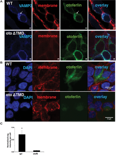
Fig. 6 Loss of the TMD reduces expression of otoferlin in cell culture. (A) Representative images of cells transfected with CFP-VAMP and either WT mouse otoferlin or ΔTMD, a mouse otoferlin with a mutation matching the otofB mutation. (B) Representative images of cells labeled with 4′,6-diamidino-2-phenylindole (blue), wheat germ agglutinin for the cell membrane (red), and otoferlin (green). (C) Quantitation of the intensity of otoferlin from transfected HEK cells (t test, p < 0.001). Scale bar for A and B = 2 µm, and 10 µm for C and D.
Acknowledgments
This image is the copyrighted work of the attributed author or publisher, and
ZFIN has permission only to display this image to its users.
Additional permissions should be obtained from the applicable author or publisher of the image.
Full text @ Mol. Biol. Cell