
|
Fig. 1
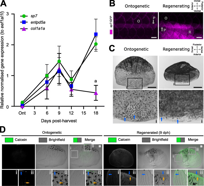
Regenerating scales express metabolic active bone growth markers at 9 days post-harvest. A Quantitative real-time PCR of osteoblast (sp7), mineralisation (entpd5a), and matrix growth (col1a1a) amplicon expression (n = 4 fish). Two-way ANOVA analysis showed that there was no interaction factor variance (p = 0.133) but that time (f(4) = 9.47, p< 0.001) and amplicon (f(2) = 4.62, p< 0.05) had independent statistically significant effects. Tukey’s multiple comparison determined that col1a1a was significantly lower expressed at 18 days post-harvest (dph) compared to other amplicons (a). B In toto fluorescent stereomicroscope images of flanks from sp7:GFP transgenic fish showing ontogenetic (O) and 9 dph regenerated scales (R) in an anterior (left) to posterior (right) direction). Note the auto-fluorescent pigment stripe (P) and the compass orientation is used to depict in toto scales in this paper with the distal (posterior) edge pointing caudally. C In situ alkaline phosphatase staining of a pre-collection and 9 dph scale with insets (i) showing anterior part of the scale. Blue arrows indicate ALP+ cells which appear to be larger and more extended in ontogenetic than regenerating scales. Compass orientation is used to depict in situ scales in this paper as the anterior region was closest to the scale dermal socket (pre-harvest). D Representative stereomicroscope images of live calcein green staining labelling newly deposited calcium phosphate in ontogenetic and regenerating scales (n = 4 fish each condition). Blue arrow indicates elevated levels of calcein green labelling compared to surrounding signal whereas orange arrows indicate small puncta of enhanced signal. Insets show lateral circuli (i) and posterior epidermal (containing pigmentating melanocytes (M) region (ii). Scale bar: 100 μm