
|
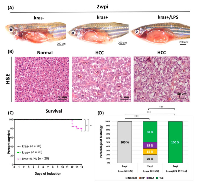
Figure 6
LPS promoted HCC progression in adult kras+ transgenic zebrafish. The wild-type (kras−) control, kras+, and kras+/LPS transgenic zebrafish were treated at 4 mpf with 10μg/mL Dox alone or with 10μg/mL Dox + 40ng/mL LPS, and samples were taken at 2 wpi. (A) The upper row shows the internal organs of the abdomen, and (B) the lower row displays H&E staining of liver sections (white dotted frame: liver). (C) Kaplan-Meier survival curves reveal the percentage of survival at 2 wpi. (D) Histological analysis shows that kras+ and kras+/LPS transgenic zebrafish developed HCC at 2 wpi. Scale bar: 50 or 200 μm. Student’s t-test or one-way ANOVA were used to assess differences between variables: * p < 0.05, *** p < 0.001.