
|
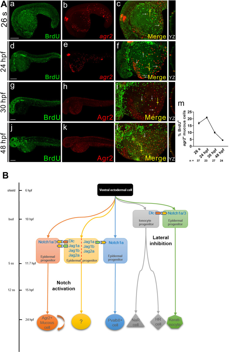
Fig 6 agr2+ EMC population is maintained by proliferation.(A). Double immunofluorescence and in situ hybridization or double immunofluorescence reveals colocalization of BrdU (green) and agr2/Agr2 (red) in the yolks of embryos at 26 s, 24, 30 and 48 hpf. BrdU labeling assay revealed that agr2+ EMCs proliferate after differentiation. The highest proliferation rate was detected at 24 hpf. White arrows indicate BrdU+agr2+ EMCs. Yellow arrows indicate positions of YZ projections of confocal images. Mean ± SEM. Scale bars, 100 μm. (B). A model depicts the timing at which sequential specification of ionocytes/keratinocytes and agr2+ EMCs/pvalb8-positive cells occurring in zebrafish embryos. The previously-explored specification of ionocytes and keratinocytes via Dlc-mediated Notch1a/3 lateral inhibition occurs at late gastrulation stage, while the newly-identified induction of agr2+ EMCs by Dlc and Jag1a/1b/2a-mediated activation of Notch1a/3 signaling and the induction of pvalb8-positive cells via Jag1a/Jag1b/Jag2a-mediated activation of Notch1a signaling occur during segmentation stage. Underlying data are available in S1 Data.