
|
FIGURE 1
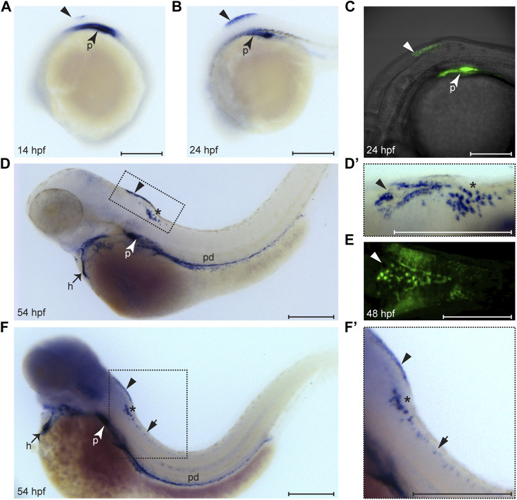
Expression pattern of wt1a during embryonic and larval development. Wt1a expression was analyzed by whole mount in situ hybridization (A, B, D, D′, F, F′) and via examination of the EGFP signal in animals carrying the wt1a:EGFP transgene (C, E). (A) In addition to the known and prominent expression in the developing pronephros (arrowhead), weak expression of wt1a is present in the presumptive dorsal hindbrain at 14 hpf (triangle). (B) The dorsal hindbrain domain appeared more prominent at 24 hpf. (C) Overlay of transmission and fluorescent images of a Tg(wt1a:EGFP) embryo reveals that the domain described in B is located at the most posterior region of the hindbrain. (D) At 54 hpf wt1a expression is detected in the pronephros, in the heart, along the pronephric duct and in two domains of the hindbrain. In addition to the above mentioned very dorsally located domain (arrowhead) the wt1a transcript is also present in a more ventral region (asterisk). (D′) A closer, dorsolateral view on both hindbrain domains illustrates that the dorsal domain (arrowhead) has a triangular shape with a compact structure in the middle. (E) The EGFP signal arising from a 2 day old embryo of the wt1a transgenic line closely recapitulates the v-shaped expression described in D’. (F-F′) Upon extended staining of a 54 hpf larva wt1a expressing cells are also detectable in the spinal cord (short arrow). A-D, F and F′ are lateral views, D′ is a dorsolateral and E a ventral view. All images are with rostral to the left. h: heart; p: pronephros; pd: pronephric duct. Scale bars: 200 μm.