
|
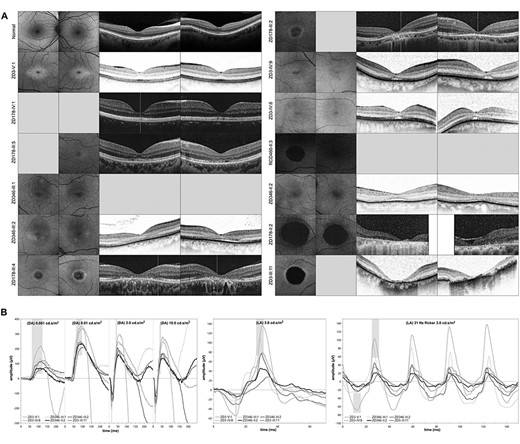
Fig. 3 Clinical presentation of patients affected by autosomal dominant cone dystrophy caused by duplications at the IRXB gene cluster: (A) Fundus autofluorescence (FAF) and optical coherence tomography (OCT) images of 13 patients with a duplication at the IRXB gene cluster, sorted by the age at examination (compare Fig. 1, Table 2 and Supplementary Material, Table S1). Note the central hypoautofluorescence and paracentral hyperautofluorescence on FAF imaging in the younger patients and the well-demarcated central hypoautofluorescence in the older patients, as well as the central thinning of the outer nuclear layer, the paracentral hyporeflectivity of the retinal pigment epithelium, and/or a subretinal cleft on OCT imaging in the younger patients and the central atrophy in the older patients. (B) Electroretinography (ERG) of six patients sorted by age, commencing with the youngest in light gray and ending with the oldest in black. The normative data are highlighted in gray bars. Under scotopic light conditions, the ERG (indicated by ‘DA’, left) shows some decline, which may be explained by an ageing effect. The photopic ERG (indicated by ‘LA’, middle: single flash; right: 30 Hz flicker) shows a decline in the amplitude of the a- and b-wave.