
|
Fig 1
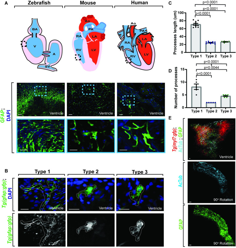
(A) Confocal maximum z-projection from adult zebrafish, mouse, and human ventricle stained with GFAP. Schematics of hearts per species (above) indicate location of imaging. Gfap+ cells are present across species. (B) Confocal 10 μm z-projection of adult Tg(gfap:gfp) ventricles with 3 distinct morphologies of gfap+ cells. (C) Quantification of process length between gfap+ cell types. (D) Quantification of process number per gfap+ cell type. (E) Confocal maximum z-projection from adult Tg(myl7:gfp) ventricles stained with AcTub and GFAP showing localization. Gfap+ cells localize with AcTub+ axons. Data are represented as mean ± SEM. Scale bar equals 10 μm. Statistics summarized in S1 Table. See S1 Data for raw data. A, atrium; AcTub, acetylated tubulin; AO, aorta; BA, bulbous arteriosus; LA, left atrium; LV, left ventricle; PA, pulmonary artery; RA, right atrium; RV, right ventricle; SV, sinoatrial valve; V, ventricle.