
Image
|
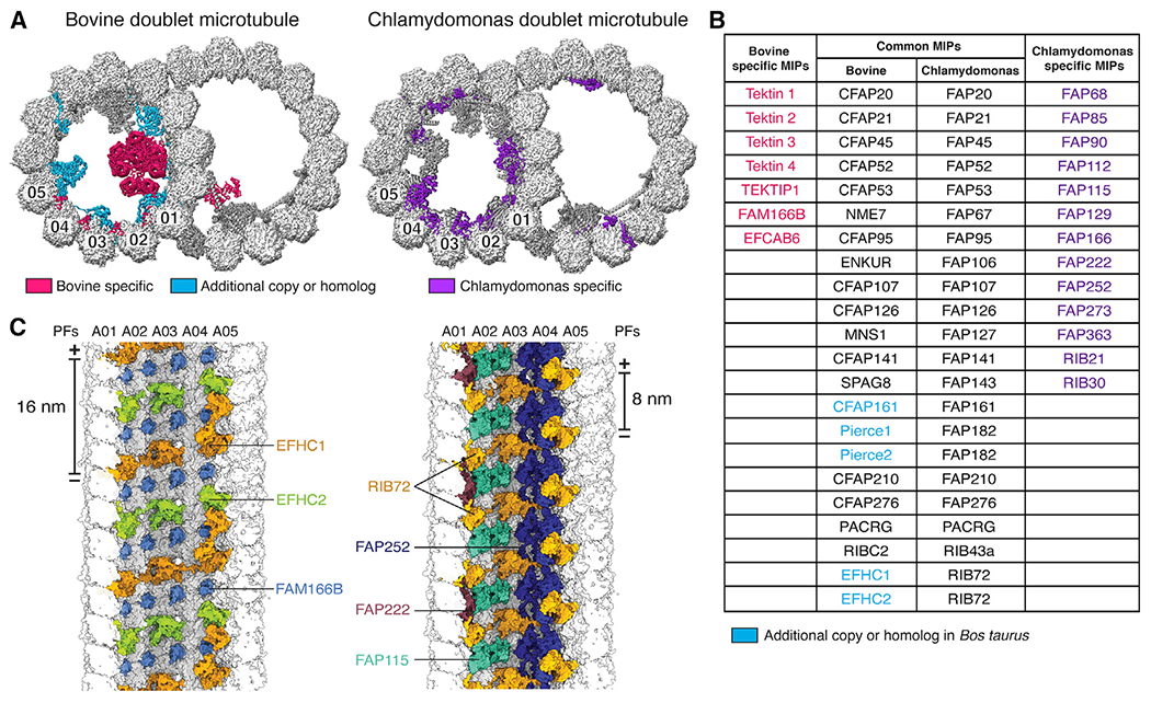
Figure Caption
Figure 2.
Microtubule inner proteins (MIPs) of mammalian DMTs
(A) Cross-sections of bovine (left) and Chlamydomonas (right) DMTs, with MIPs colored by conservation. MIPs present in both organisms are colored dark gray.
(B) Table of MIPs identified in bovine and Chlamydomonas DMTs. Protein names are colored according to their classification in (A). The names of shared MIPs are given for bovine and C. reinhardtii.
(C) View from the A-tubule lumen showing bovine (left) and Chlamydomonas (right) MIPs bound to PFs A01–A05. Chlamydomonas RIB72 repeats every 8 nm, whereas its two bovine paralogs, EFHC1 and EFHC2, repeat every 16 nm.
See also Video S1.
Acknowledgments
This image is the copyrighted work of the attributed author or publisher, and
ZFIN has permission only to display this image to its users.
Additional permissions should be obtained from the applicable author or publisher of the image.
Reprinted from Cell, 184(23), Gui, M., Farley, H., Anujan, P., Anderson, J.R., Maxwell, D.W., Whitchurch, J.B., Botsch, J.J., Qiu, T., Meleppattu, S., Singh, S.K., Zhang, Q., Thompson, J., Lucas, J.S., Bingle, C.D., Norris, D.P., Roy, S., Brown, A., De novo identification of mammalian ciliary motility proteins using cryo-EM, 5791-5806.e19, Copyright (2021) with permission from Elsevier. Full text @ Cell