
|
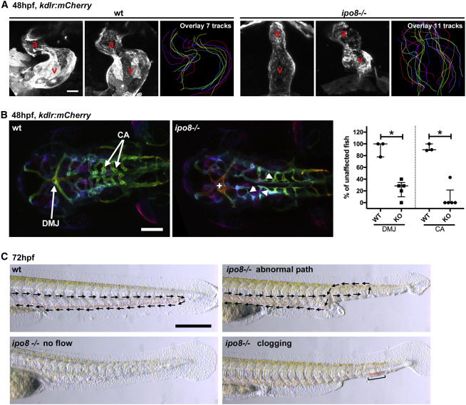
Fig. 4 Ipo8 deficiency causes cardiovascular defects in zebrafish (A) Defects in heart chamber formation exemplified by two maximal projections of confocal Z stacks of the hearts of WT and ipo8−/− 48 hpf embryos carrying the kdrl:mCherry transgene to label endothelial cells. For each line, the third panel shows the overlays of the outlines of the 7 (WT) and 11 (ipo8−/−) analyzed hearts. a, atrium; v, ventricle. Scale bar represents 30 μm. (B) Maximal projections of depth color-coded confocal Z stacks of the head vessels of WT and ipo8−/− embryos at 48 hpf, and quantifications of the percentage of fish showing dorsal midline junction (DMJ) defects and central arteries (CA) differentiation defects. Twenty-three WT and 30 ipo8−/− embryos from 3 and 5 clutches, respectively, were analyzed. Median and IQR are shown. p values were calculated by Mann Whitney test (∗p < 0.05). +, abnormal DMJ; arrowheads, abnormal CA. Scale bar represents 100 μm. (C) Maximal projections of time-lapses of Nomarsky imaging of the tails of 3 dpf larvae highlighting absence of blood circulation (no flow) or defects in blood vessel patterning (abnormal path and clogging) in ipo8−/− mutants. Arrows indicate flow direction. Scale bar represents 200 μm.