
|
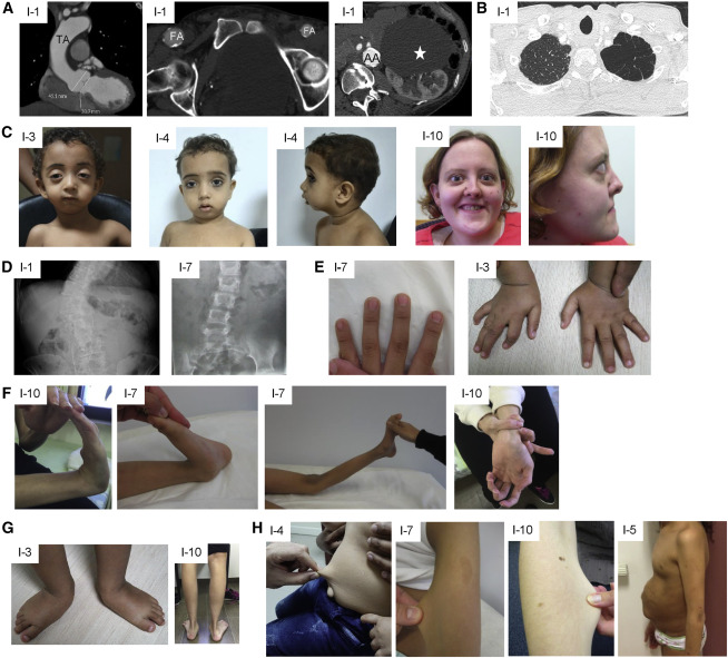
Fig. 1 Clinical features of affected individuals (A) CT scan showing dilated aortic root and thoracic aorta (TA), calcified asymmetric large femoral arteries (FA), calcified and dilated abdominal aorta (AA), and large renal cyst (white star) in individual 1. (B) CT scan showing emphysema of the apex of the left lung in individual 1. (C) Proptosis, micrognathia, and hypertelorism in individuals 3, 4, and 10. (D) X-ray showing scoliosis in individuals 1 and 7. (E) Arachnodactyly in individuals 3 and 7. (F) Hyperlaxity of small and large joints in individuals 10 and 7. (G) Pes planus and talipes equinovarus in individuals 10 and 3, respectively. (H) Skin hyperextensibility in individuals 4, 7, and 10 and umbilical hernia in individual 5.