
Image
|
Figure Caption
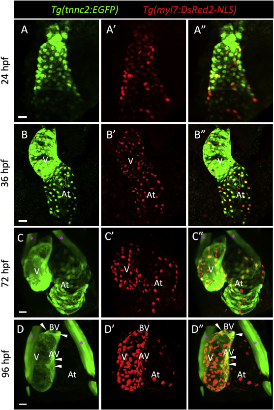
Fig. 2 The tnnc2 reporter is expressed by embryonic cardiomyocytes in zebrafish. (A–D) Confocal images (maximum intensity projections) of wholemount Tg(tnnc2:EGFP); Tg(myl7:DsRed2-NLS) hearts at 24 (A-A″), 36 (B–B″), 72 (C–C″) and 96 (D-D″) hpf. White arrowheads point to EGFP+ CMs around the atrioventricular (AV) and bulboventricular (BV) areas at 96 hpf (D–D″). Magenta asterisks mark craniofacial muscles. V: ventricle; At: atrium. Scale bars: 20 μm.
Acknowledgments
This image is the copyrighted work of the attributed author or publisher, and
ZFIN has permission only to display this image to its users.
Additional permissions should be obtained from the applicable author or publisher of the image.
Reprinted from Developmental Biology, 476, Tsedeke, A.T., Allanki, S., Gentile, A., Jimenez-Amilburu, V., Rasouli, S.J., Guenther, S., Lai, S.L., Stainier, D.Y.R., Marín-Juez, R., Cardiomyocyte heterogeneity during zebrafish development and regeneration, 259-271, Copyright (2021) with permission from Elsevier. Full text @ Dev. Biol.