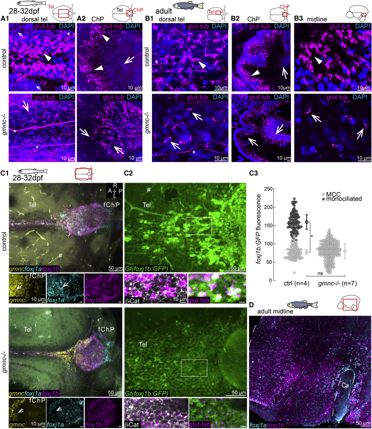
IMAGE

Figure 4

ID
ZDB-IMAGE-211029-104
Genes
Source
Figures for D'Gama et al., 2021
Image
Figure Caption

Figure 4

gmnc is required for multiciliation in ependymal cells

(A and B) Absence of MCCs in 1-month-old (A1 and A2; n = 3 ctrl; 3 mutants) and adult brains of gmnc mutants (B1–B3; n = 3 ctrl; 3 mutants). Arrowheads show MCC in controls. All ciliated cells in gmnc mutant are monociliated (arrow). (A1) 1-month dorsal telencephalon and (A2) ChP. (B1) Adult dorsal telencephalon. (B2) Anterior portion of the adult ChP. (B3) Adult midline above the Ca.

(C1–C3) gmnc enhances the expression of foxj1a and foxj1b in the TC and ChP. (C1) HCR revealed a gmnc-dependent foxj1a expression, particularly in the anterior ChP (arrow in insets). n = 4 controls; 6 mutants. The hashtag symbol shows nonspecific signal in blood vessels. (C2) foxj1b:GFP remained expressed in gmnc mutant with reduced and more homogeneous levels (n = 4 controls; 7 mutants). β-catenin and glutamylated tubulin staining was used for quantification of GFP levels in C3. (C3) Monociliated cells express less foxj1b:GFP than MCCs and monociliated cells in gmnc mutant express similar foxj1b:GFP levels as control monociliated cells. All data points are plotted; mean and SD of n fish; p < 0.05 calculated on the average fluorescence per fish using rank sum test.

(D) HCR shows that foxj1a and foxj1b remained expressed in the telencephalic midline of gmnc mutant (n = 3).

Asterisk indicates axons. A, anterior; P, posterior; R, right; L, left; Tel, telencephalon.

See also Figure S3.

Figure Data
Acknowledgments
This image is the copyrighted work of the attributed author or publisher, and ZFIN has permission only to display this image to its users. Additional permissions should be obtained from the applicable author or publisher of the image. Full text @ Cell Rep.