IMAGE

Fig. 1.

ID
ZDB-IMAGE-211009-52
Source
Figures for Honkoop et al., 2021
Image
Figure Caption

Fig. 1.

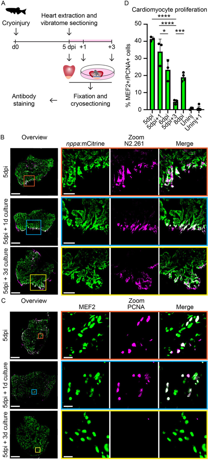
Cardiac slices retain characteristics of the regenerating heart. (A) Schematic representation of workflow. (B) Immunolabeling on 5 dpi whole hearts, 5 dpi cardiac slices after 1 day culture and 5 dpi cardiac slices after 3 day culture from a Tg(nppa:mCitrine) transgenic line. Trabecular cardiomyocytes are marked with mCitrine (green) and embryonic myosin is marked with N2.261 (magenta). Scale bars: 200 µm (overview); 50 µm (zoom). (C) Immunolabeling on 5 dpi whole hearts, 5 dpi cardiac slices after 1 day culture and 5 dpi cardiac slices after 3 day culture from a Tg(nppa:mCitrine) transgenic line. Cardiomyocyte nuclei are marked with MEF2 (green) and proliferating nuclei are marked with PCNA (magenta). Scale bars: 200 µm (overview) or 10 µm (zoom). (D) Quantification of MEF2+/PCNA+ cells in the border zone of injured hearts/cardiac slices and remote zone of uninjured hearts/cardiac slices. Each dot represents a result from one heart or cardiac slice. Data are mean±s.d. *P<0.05, ***P<0.001, ****P<0.0001.

Acknowledgments
This image is the copyrighted work of the attributed author or publisher, and ZFIN has permission only to display this image to its users. Additional permissions should be obtained from the applicable author or publisher of the image. Full text @ Development