
Image
|
Figure Caption
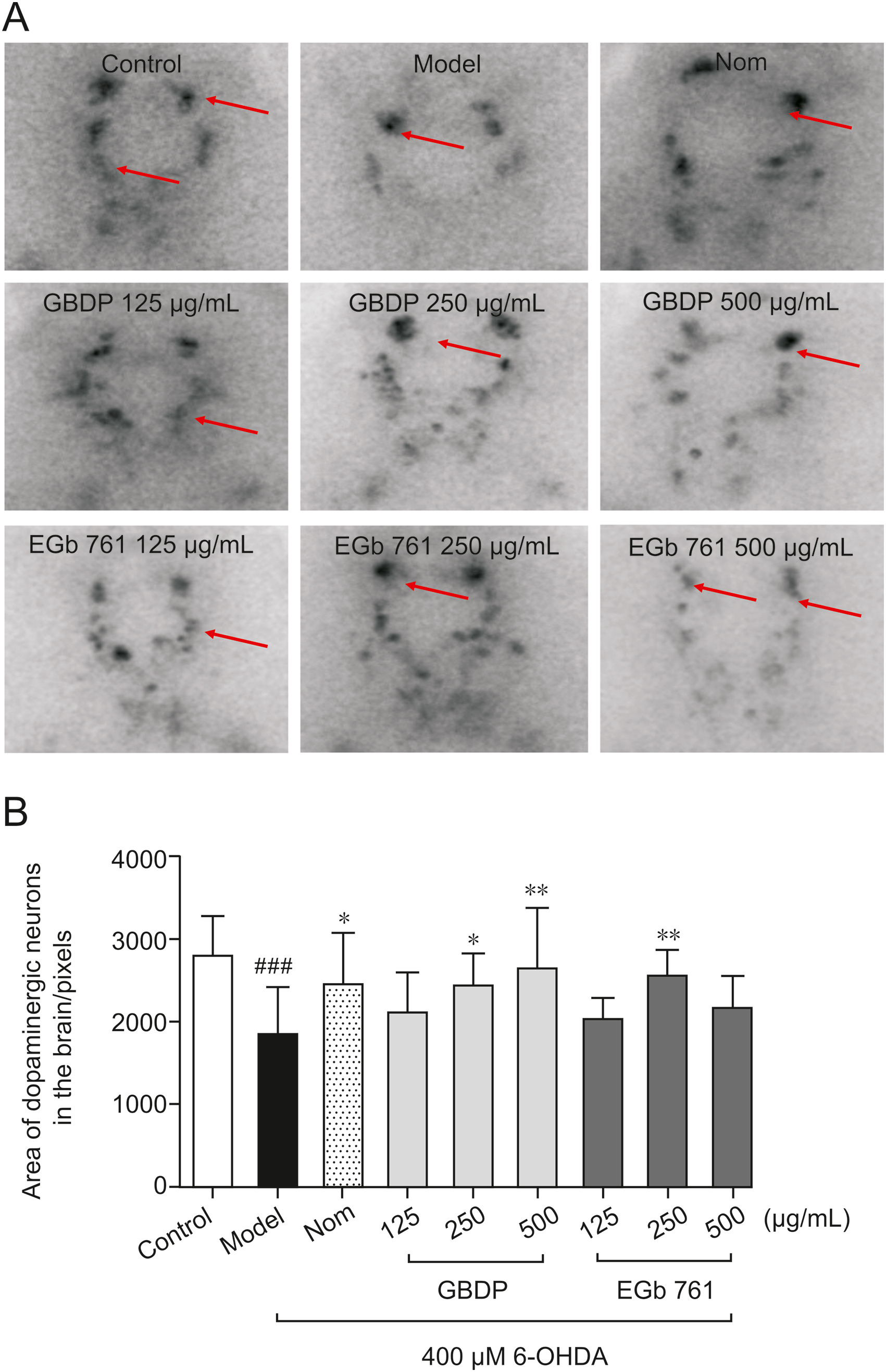
Fig. 3 GBDP protected against 6-OHDA-induced dopaminergic neuron loss in zebrafish. (A) Representative images of DA neurons in the zebrafish brain, indicated by tyrosine hydroxylase immunostaining. Red arrow: dopaminergic neurons in the zebrafish brain. (B) The area of the dopaminergic neurons calculated for each group. Data were analyzed by one-way ANOVA followed by Dunnett’s test. ###P < 0.0001, compared with the control group; ∗P < 0.05, ∗∗P < 0.001 compared with the 6-OHDA group. n = 10 per group.
Figure Data
Acknowledgments
This image is the copyrighted work of the attributed author or publisher, and
ZFIN has permission only to display this image to its users.
Additional permissions should be obtained from the applicable author or publisher of the image.
Full text @ J Pharm Anal