
|
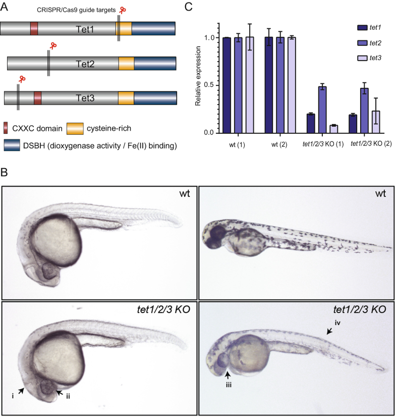
Fig. 1 Phenotypes of tet1/2/3 KO F0 zebrafish following CRISPR/Cas9 microinjections. (a) Zebrafish Tet protein structure and guide RNA design. (b) Top left: uninjected 24 hpf zebrafish, bottom left: tet1/2/3 KO injected 24 hpf zebrafish displaying improperly formed midbrain hindbrain boundary (i) and microphthalmia (ii). Top right: uninjected 48 hpf zebrafish; bottom right: tet1/2/3 KO injected 48 hpf zebrafish displaying microphthalmia (iii), altered pigmentation and curved trunk (iv). (c) qPCR quantification of two biological replicates of uninjected wild type (wt) and injected (tet1/2/3 KO) F0 zebrafish. Each gene is tested in triplicate and represented as the mean relative expression to wt. Error bars represent standard deviation