
Image
|
Figure Caption
Figure 8
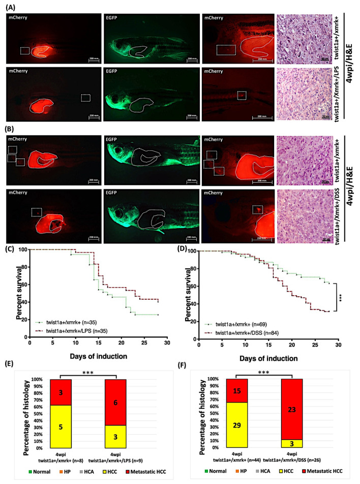
Figure 8. Induction of tumor metastasis in twist1a+/xmrk+ transgenic zebrafish via Dox and 4-OHT treatment under exposure to LPS or DSS. Twist1a+/xmrk+ transgenic zebrafish were treated with 60 μg/mL Dox and 1 μg/mL 4-OHT starting from 4 mpf with sampling performed at 4 wpi. (A,B) Immunofluorescence analysis of twist1a+/xmrk+ liver tumor metastasis at 4 wpi with corresponding H&E histological examination of liver sections. Scale bar: 50 or 200 μm. (C,D) Kaplan–Meier survival curves showing days post-induction plotted against percentage survival to 4 wpi. (E,F) Histological examination confirmed that at 4 wpi, all twist1a+/xmrk+ transgenic zebrafish developed HCC or metastatic HCC. Differences among the variables were assessed using one-way ANOVA. Statistical significance: *** p < 0.001.
Figure Data
Acknowledgments
This image is the copyrighted work of the attributed author or publisher, and
ZFIN has permission only to display this image to its users.
Additional permissions should be obtained from the applicable author or publisher of the image.
Full text @ Pharmaceuticals (Basel)