
Image
|
Figure Caption
Figure 5
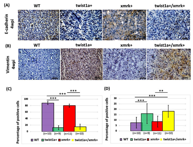
Figure 5. Assessment of E-cadherin and vimentin staining in instances of HCC progression in twist1a+/xmrk+ double transgenic zebrafish. Immunohistochemical staining was performed on liver paraffin sections from WT, twist1a+, xmrk+, and twist1a+/xmrk+ zebrafish at 4 wpi. Staining of markers of EMT activation: (A) E-cadherin and (B) vimentin. Scale bar: 50 μm. Quantification of the percentage of cells testing positive for (C) E-cadherin and (D) vimentin. Differences among the variables were assessed using Student’s t-tests. Statistical significance: ** p < 0.01, *** p < 0.001.
Figure Data
Acknowledgments
This image is the copyrighted work of the attributed author or publisher, and
ZFIN has permission only to display this image to its users.
Additional permissions should be obtained from the applicable author or publisher of the image.
Full text @ Pharmaceuticals (Basel)