
|
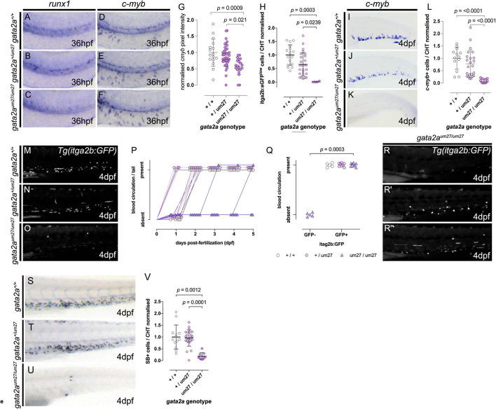
FIGURE 2 Impaired HSPC development in gata2a mutants. (A–C) Expression of runx1 by in situ hybridization in 36 hpf embryos, showing decreased expression in gata2aum27/um27 homozygotes. (D–F) Lateral views of c-myb WISH on 36 hpf Gata2aum27 mutants showing decreased expression in in gata2aum27/um27 homozygotes. (G) Quantification of c-myb expression from in situ hybridization images in the AGM of 36 hpf embryos. In (H) mutant fish carrying itga2b:GFP transgene, labeling HSPCs that reside in the CHT, were used to quantify GFPlow cells in the CHT of 56 hpf embryos, showing an allele dependent decrease in GFPlow cells. (I–K) Lateral views of 4 dpf larvae showing the expression of c-myb by in situ hybridization. (L) Quantification of c-myb+ cells in the CHT of 4 dpf mutant larvae. (M–O) Lateral views of the tails of 4 dpf mutant Tg(itga2b:GFP) fish showing a decrease of GFP+ cells in gata2aum27/um27 homozygotes. In (P), the presence (up) or absence (bottom) of blood flow in the tail of Gata2aum27 mutant fish was monitored each day until 5 dpf. Notice the gata2aum27/um27 homozygotes (purple triangles) gaining blood circulation at 3 and 5 dpf. (Q) Plot showing the correlation of the presence (up) or absence (bottom) of blood flow in the tail of Gata2aum27 mutant fish and the presence (right) or absence (left) of Tg(itga2b:GFP)+ cells in 4 dpf fish. At 4 dpf, some gata2aum27/um27 homozygotes (purple triangles) show GFP+ cells, all of which also exhibit circulation in the tail. (R–R″) Lateral views of 4 dpf mutant Tg(itga2b:GFP) fish showing a range of phenotypes of gata2aum27/um27 homozygotes. (S–U) Lateral views of 4 dpf mutant larvae stained with SB. (V) Quantification of SB+ cells in the CHT of 4 dpf larvae shows decreased number of granulocytes in gata2aum27/um27 homozygotes.