
|
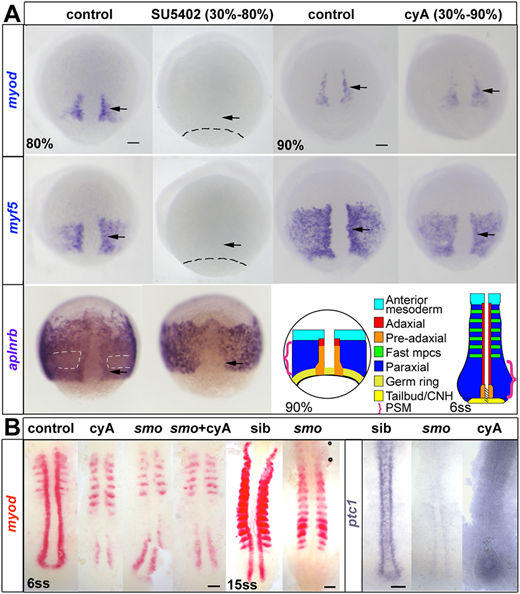
Fig. 1 ISH for myod and myf5 in control untreated, cyA-treated (100 µM) and SU5402-treated (60 μM) wild-type or mutant embryos, shown in dorsal view, anterior to top. (A) Adaxial (arrows) and paraxial myod and myf5 mRNAs are lost upon SU5402 treatment from 30% to 80 or 90% epiboly (dashes indicate approximate position of the germ ring) but are unaffected by cyA treatment. The anterior mesoderm marker aplnrb is normally downregulated in paraxial presomitic cells expressing myf5 (white dashes) and upregulated in adaxial cells (arrows). Both changes were absent after SU5402 treatment. Schematics illustrate the location of equivalent cell types at two successive stages. CNH, chordoneural hinge (hatched); mpcs, muscle precursor cells; PSM, presomitic mesoderm (brackets). (B) Smob641 mutants retain pre-adaxial myod mRNA at 6ss even after cyA treatment, but lack pre-adaxial myod mRNA at 15ss. ptc1 (ptch2) mRNA downregulation shows that both smo mutation and cyA treatment (shown after longer colour reaction) fully block Hh signalling throughout the axis. Scale bars: 50 µm.