
|
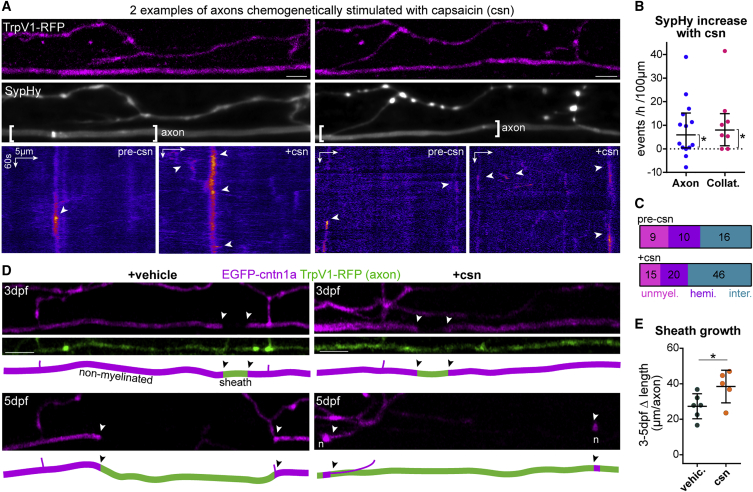
Figure 6
Chemogenetic stimulation of vesicular fusion promotes sheath growth
(A) Two examples of SypHy activity in TrpV1-tagRFP+ reticulospinal axons before (pre-csn) and during capsaicin treatment (+csn).
(B) Capsaicin increased axonal and collateral SypHy in TrpV1+ neurons above baseline (difference from zero: p = 0.013 axonal, p = 0.031 collateral, Wilcoxon signed-rank test, 14 axons and 8 collaterals from 14 and 8 animals, respectively).
(C) Axonal event distribution before (n = 35 events) and after (n = 81 events) csn treatment.
(D) Single TrpV1+ axons treated with vehicle or capsaicin throughout myelination (arrowheads, putative heminodes; n, putative nodes of Ranvier).
(E) Stimulation promotes sheath growth (3–5 dpf change: p = 0.04, Student’s t test). 6 control axons and 5 stimulated axons from 6 and 5 animals, respectively.
Scale bars, 5 μm (A), 10 μm (D), 60 s (A). Graphs in (B) shows median and interquartile range and graphs in (E) display mean and standard deviation.
See also