
|
Figure 4
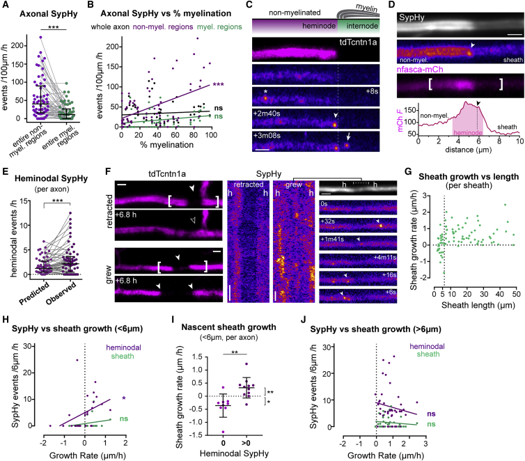
Axonal vesicular fusion is enriched at heminodes during nascent sheath growth
(A) Within myelinated axons, SypHy is more frequent in non-myelinated regions (p < 0.0001, Wilcoxon matched-pairs signed-rank test, 58 myelinated axons from 53 animals).
(B) Percent myelination correlates with SypHy frequency in non-myelinated regions (Pearson’s r, 0.46, p < 0.0001; in myelinated regions r, 0.25, p = 0.05; in whole axon r, 0.10, p = 0.43).
(C) SypHy activity near a putative heminode, annotations as in
(D) nfasca-mCherry is enriched at putative heminodes (arrowhead, heminodal SypHy event). Graph indicates mCherry fluorescence intensity profile (in a.u.) along bracketed region.
(E) Observed heminodal Syphy frequency is higher than predicted if events in non-myelinated regions were uniformly distributed (p = 0.0004, Wilcoxon matched-pairs rank test, 58 axons from 53 animals).
(F) tdTcntn1a profiles and SypHy activity in retracting and growing nascent sheaths (h = heminode).
(G) Relation between sheath length and growth rate, note sheaths <6 μm may grow or shrink, but >6 μm sheaths mostly grow (85 sheaths from 39 axons in 35 fish).
(H) Nascent sheath growth rate correlates with heminodal SypHy frequency (Pearson’s r: 0.39, p = 0.043) but not under the sheath (Pearson’s r: 0.31, p = 0.111). 85 sheaths from 39 axons from 36 animals.
(I) Nascent sheaths with heminodal SypHy activity grow faster than those without (p = 0.001 with versus without, Mann-Whitney test; p = 0.01 and p = 0.039 non-zero growth rate with and without heminodal SypHy, one-sample Wilcoxon test; 23 sheaths in N = 14 axons from 13 animals).
(J) Growth rate of sheaths >6 μm does not correlate with SypHy activity (heminodal Pearson’s r: −0.14, p = 0.381; sheath Pearson’s r: −0.12, p = 0.441).
Scale bars, 2 μm (C, D, and F), 60 s (F). Graphs display median and interquartile range.
See also