|
Fig. 5
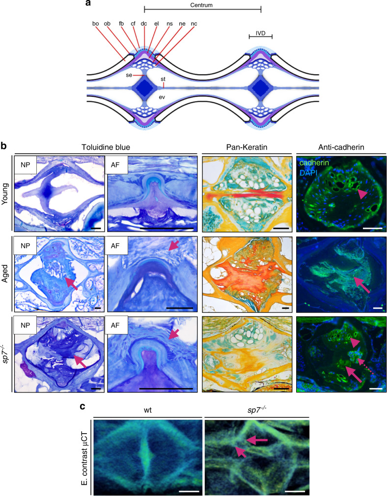
IVDD histopathology underlying 3D disc changes in zebrafish.

|
|
Fig. 5
IVDD histopathology underlying 3D disc changes in zebrafish.