
Image
|
Figure Caption
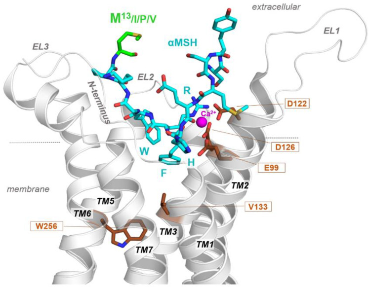
Figure 6
Molecular model of α-MSH and zebrafish MC1R complex. The MC1R (carton representation) binds α-MSH in a cavity between the extracellular loops (ELs) and the transmembrane helices (TMs). Visualized are amino acids (sticks) covering the ligand binding site and are highly important for ligand and co-factor calcium (Ca2+) binding. All α-MSH ligand variants from different species share a conserved core motif (HRFW) involved in receptor-ligand contacts. The α-MSH C-terminus with position 13 (M13) is not in direct contact with the receptor as supposed by MC4R/α-MSH or MC1R/α-MSH complexes.
Acknowledgments
This image is the copyrighted work of the attributed author or publisher, and
ZFIN has permission only to display this image to its users.
Additional permissions should be obtained from the applicable author or publisher of the image.
Full text @ Int. J. Mol. Sci.