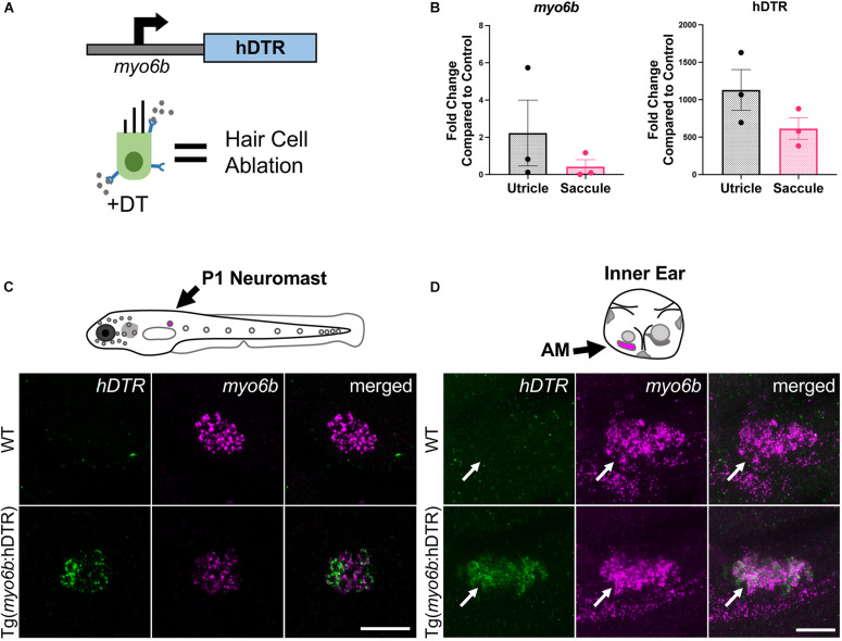
IMAGE

FIGURE 1

ID
ZDB-IMAGE-210909-6
Source
Figures for Jimenez et al., 2021
Image
Figure Caption

FIGURE 1

The Tg(myo6b:hDTR) zebrafish. (A) Schematic representation of the human diphtheria toxin receptor (hDTR) construct driven by the hair cell-specific myo6b promoter and representation of the hair cell-specific ablation approach. Diphtheria toxin (DT) is internalized in cells specifically expressing the human version of the DT receptor, triggering cell death. (B) qRT-PCR analysis on untreated Tg(myo6b:hDTR) zebrafish saccule and utricle showing expression of hDTR and myo6b relative to wild-type (WT) animals. The values are represented as the mean ± SEM from three independent samples. (C) Schematic depicts a lateral view of a whole zebrafish larva with neuromasts (gray circles). The P1 neuromast examined for imaging is indicated by magenta fill. In situ HCR using probes targeting hDTR and myo6b in hair cells of a single P1 neuromast of 3 dpf WT and Tg(myo6b:hDTR) embryos is shown. Brightness and contrast adjusted 50 and 25%, respectively. Scale bar 20 μm. (D) Schematic depicts the left ear of a larval zebrafish with anterior macula indicated by magenta fill. Lateral views of inner ears are shown following HCR in situ with probes targeting hDTR and myo6b in 3 dpf WT and Tg(myo6b:hDTR) embryos. Brightness and contrast adjusted 65 and 35%, respectively. Scale bar 20 μm.

Acknowledgments
This image is the copyrighted work of the attributed author or publisher, and ZFIN has permission only to display this image to its users. Additional permissions should be obtained from the applicable author or publisher of the image. Full text @ Front. Cell. Neurosci.