
|
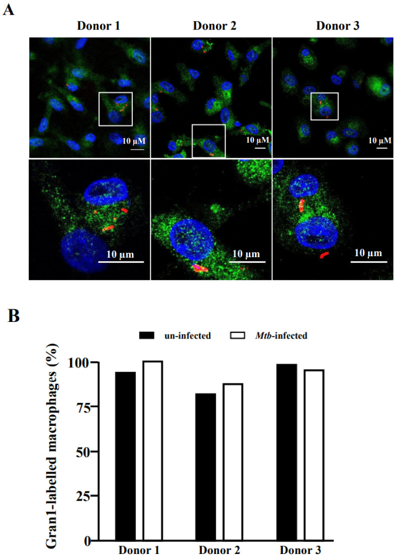
Figure 6
Uptake of Gran1 by Mtb-infected macrophages: (A) Macrophages were infected with NHS-AlexaFluor 647-labelled Mtb for 16 h, followed by 2 h incubation with Gran1. Gran1 was labelled using an anti-Gran1 antibody and a Cy2-labelled secondary antibody (green). Cell nuclei were stained with DAPI (blue). Images were acquired using an inverted laser scanning confocal microscope (Zeiss LSM 710). Images show representative areas for three donors. Original magnification x63 (upper three panels) with a 3.5-fold zoom on the region of interest (lower three panels). (B) The graph shows the numberof Gran1-labelled macrophages (%) in un-infected (black) and infected (white) cells for three independent donors.