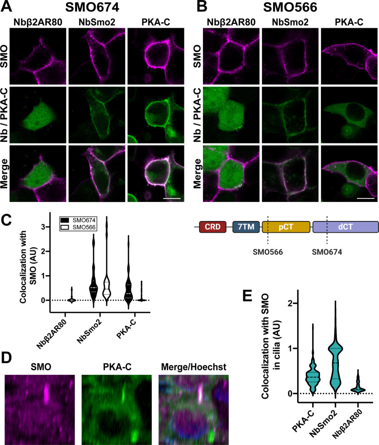
IMAGE

Fig 2

ID
ZDB-IMAGE-210826-2
Source
Figures for Arveseth et al., 2021
Image
Figure Caption

Fig 2 SMO uses its essential pCT domain to recruit PKA-C to the membrane.

HEK293 cells expressing (A) FLAG-tagged SMO674 that contains the pCT or (B) FLAG-tagged SMO566 that lacks the pCT (see cartoon at lower right) were cotransfected with Nbβ2AR80-GFP, NbSmo2-YFP, or PKA-C-YFP. Confocal microscopy images show SMO (magenta) and co-expressed proteins (green). Scale bar = 10 μm. (C) Quantification of colocalization for studies in (A) and (B). (n = 29–121 cells per condition). Note that Nbβ2AR80 displayed a background-subtracted colocalization index of “0” in all SMO674-expressing cells examined. (D) Three-dimensional reconstruction of IMCD3 cells stably coexpressing FLAG-tagged SMO (magenta) with mNeonGreen-tagged PKA-C (green). After treatment with SAG21k to induce SMO accumulation in cilia, live cells were stained with anti-FLAG antibodies (magenta) and Hoechst (blue), then examined via confocal microscopy. See S3 Fig for images from cells expressing FLAG-tagged SMO with mNeonGreen-tagged NbSmo2 or Nbβ2AR80. (E) Quantification of colocalization between SMO and PKA-C, NbSmo2, or Nbβ2AR80 for the experiment described in (D) (n = 197–236 cilia per condition). The underlying data for this figure can be found under S2 Data. dCT, distal segment of the cytoplasmic tail; pCT, proximal segment of the cytoplasmic tail; PKA-C, PKA catalytic subunits; SMO, Smoothened.

Acknowledgments
This image is the copyrighted work of the attributed author or publisher, and ZFIN has permission only to display this image to its users. Additional permissions should be obtained from the applicable author or publisher of the image. Full text @ PLoS Biol.