
|
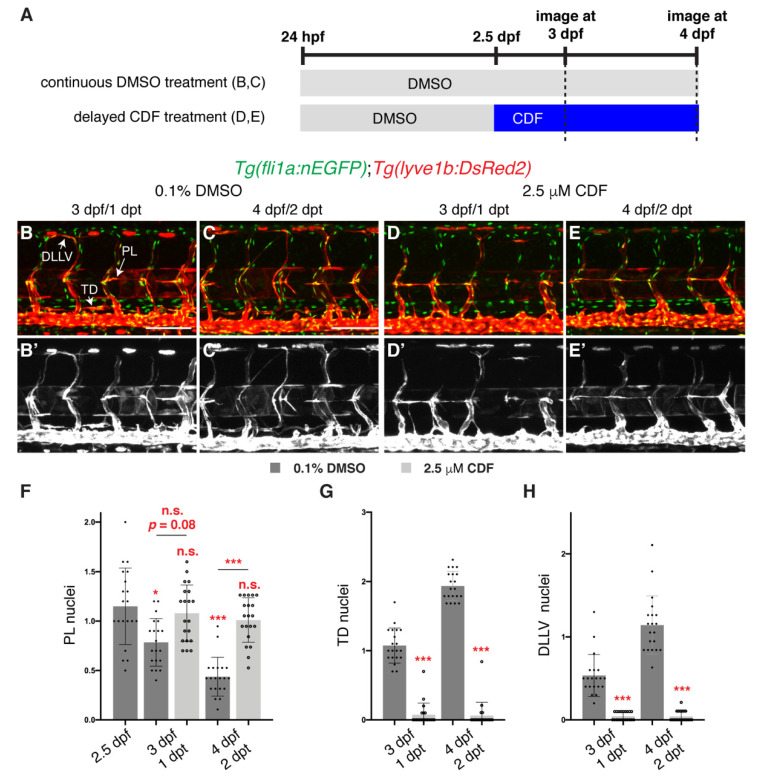
Figure 2 3,4-Difluorobenzocurcumin treatment inhibits lymphatic migration. (A) Schematic representation of the treatment schedule for larvae in images (B–E’). (B–E’) Lateral confocal images of 3 (1 days post-treatment (dpt), B,B’,D,D’) and 4 (2 dpt, C,C’,E,E’) dpf Tg(fli1a:nEGFP);Tg(-5.2lyve1b:DsRed2) larvae treated from 2.5 dpf with either 0.1% DMSO (B–C’) or 2.5 μM 3,4-Difluorobenzocurcumin (CDF, D-E’). LEC migration is stalled in larvae treated with CDF. Images (B’–E’) represent the Tg(-5.2lyve1b:DsRed2) expression of images (B–E). (F–H) Quantification of parachordal LECs (PL, F), thoracic duct (TD, G) or dorsal longitudinal lymphatic vessel (DLLV, H) nuclei per somite in Tg(fli1a:nEGFP);Tg(-5.2lyve1b:DsRed2) larvae treated with either 0.1% DMSO or 2.5 μM CDF at indicated timepoints (n = 20 embryos/larvae). Statistical test: Kruskal-Wallis test was conducted for graph F and Mann-Whitney test were conducted for graphs G and H. p ≤ 0.001 (***), p ≤ 0.05 (*), n.s. indicates not significant. Scale bars: 100 μm.