IMAGE

FIG 4

ID
ZDB-IMAGE-210726-14
Source
Figures for Horton et al., 2021
Image
Figure Caption

FIG 4

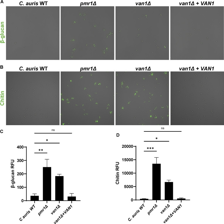
C. aurispmr1Δ and van1Δ strains display increased cell surface PAMPs. (A) Cell surface β-glucan was labeled using Fc:dectin-1 protein with Alexa Fluor 488-conjugated anti-human IgG Fc antibody and imaged by fluorescence microscopy. (B) Cell surface chitin was labeled with wheat germ agglutinin conjugated to fluorescein isothiocyanate (WGA-FITC) and assessed by fluorescence microscopy. (C and D) Total surface β-glucan and chitin were quantified by plate reader measurements of fluorescence, n = 3 mean with SEM shown, *, P < 0.05; **, P < 0.01; ***, P < 0.001 by one-way ANOVA with Holm-Sidak multiple comparisons to C. auris WT; ns, not significant.

Acknowledgments
This image is the copyrighted work of the attributed author or publisher, and ZFIN has permission only to display this image to its users. Additional permissions should be obtained from the applicable author or publisher of the image. Full text @ mSphere