IMAGE

Figure 3

ID
ZDB-IMAGE-210708-26
Genes
Antibodies
Source
Figures for Mikdache et al., 2021
Image
Figure Caption

Figure 3

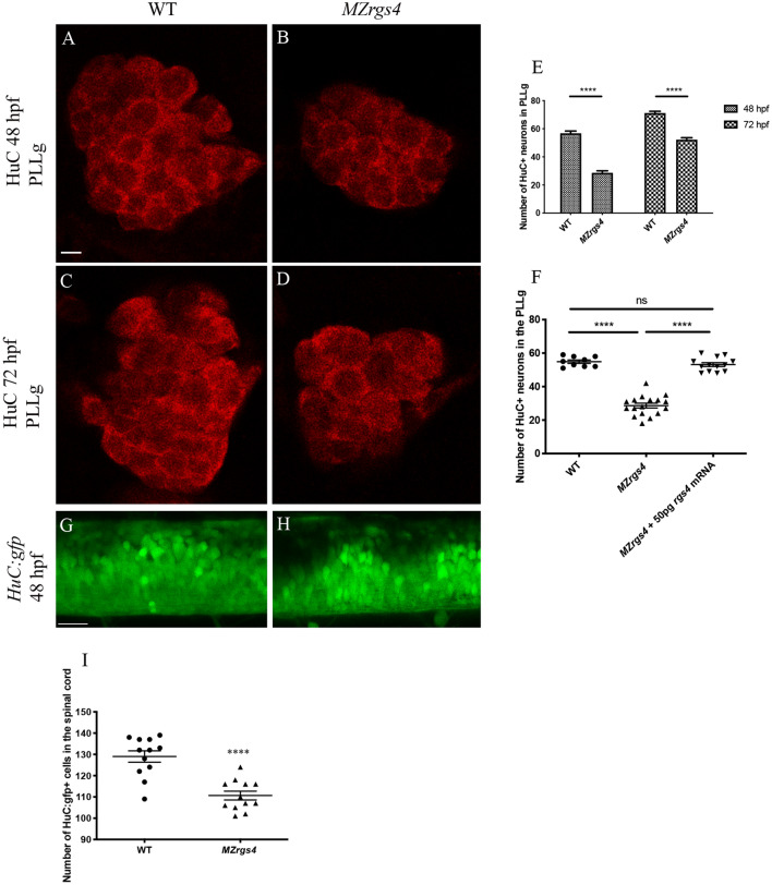
Rgs4 is required for neuronal development in the PLLg and spinal cord. (AD), single planes of confocal microscopy images representing the PLLg following HuC labelling at 48 and 72 hpf in WT (A,C) and MZrgs4 (B,D). Scale bars = 5 μm. (E) Graph showing the number of neurons within the PLLg in WT (average of 56.70 ± 1.43, n = 10 at 48 hpf; average of 71.17 ± 1.25, n = 12 at 72 hpf) and MZrgs4 embryos (average of 29.14 ± 1.71, n = 14 at 48 hpf; average of 52.14 ± 1.33, n = 21 at 72 hpf) at 48 and 72 hpf. (F) Quantification of the number of neurons in the PLLg following rgs4 mRNA injection in MZrgs4 mutants at 48 hpf (average of 54.89 ± 1.01 neurons in WT, n = 9; average of 28.65 ± 1.43 neurons in MZrgs4, n = 17; average of 53.15 ± 1.13 neurons in MZrgs4 + rgs4mRNA, n = 13). (G,H) single planes of confocal microscopy images of Tg(HuC:gfp) (G) and Tg(HuC:gfp)/MZrgs4 (H) at 48 hpf. Scale bar = 20 μm. (I) Quantification of the number of HuC:gfp + cells within the same area of the spinal cord in WT (average of 129 ± 2.70 gfp + cells, n = 12) and MZrgs4 (average of 110.7 ± 2.07 gfp + cells, n = 12) at 48 hpf.

Figure Data
Acknowledgments
This image is the copyrighted work of the attributed author or publisher, and ZFIN has permission only to display this image to its users. Additional permissions should be obtained from the applicable author or publisher of the image. Full text @ Sci. Rep.