
|
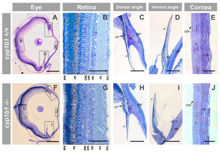
Figure 6 Ocular histology of adult (seven months) cyp1b1-KO zebrafish with Ph2 craniofacial alterations (F3). (A–J) Semithin (500 nm) tissue sections were stained with toluidine blue. The squares and rectangles indicate the areas of the images magnified in the indicated panels. No significant differences were observed between the eyes of wildtype and cyp1b1-KO zebrafish siblings. Scale bar in (A,F): 500 μm. Scale bar in (B–D,G–I): 100 μm. Scale bar in (E,J): 25 μm. RPE: retinal pigment epithelium; PR: photoreceptors; ONL: outer nuclear layer; OPL: outer plexiform layer; INL: inner nuclear layer; IPL: inner plexiform layer; GCL: ganglion cell layer; CE: ciliary epithelium; AL: annular ligament; CEP: corneal epithelium; CEN: corneal endothelium; S: stroma. The images are representative of the results observed in three fishes of each genotype. Three tissue sections per eye were analyzed.