
|
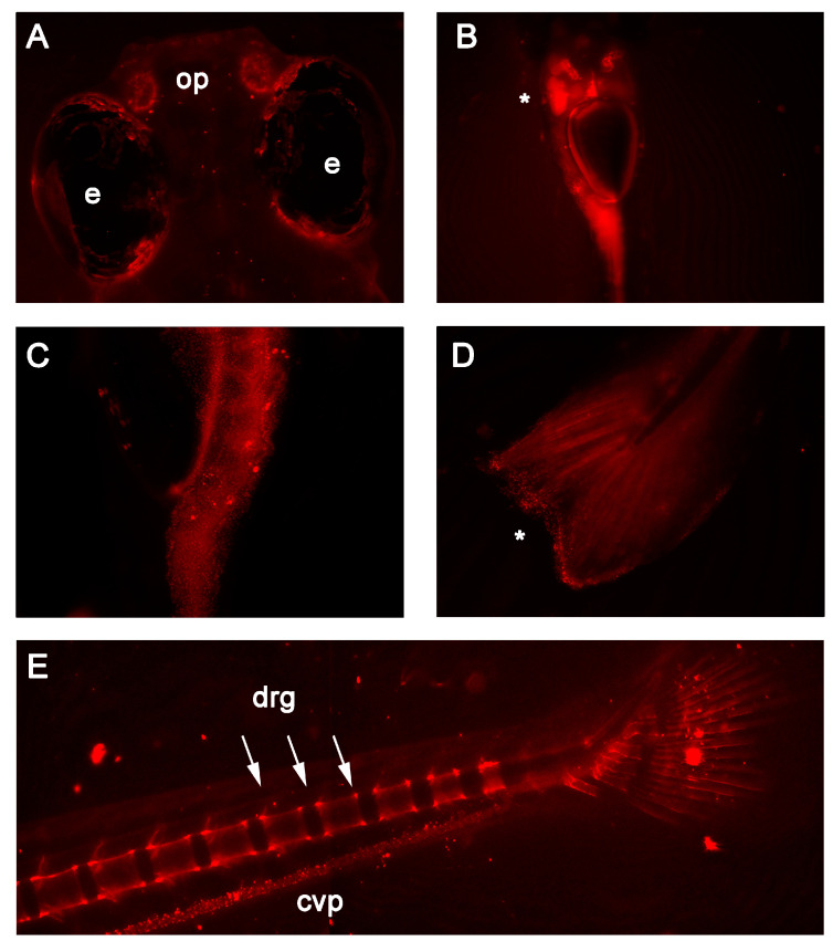
Figure 2 In vivo fluorescence detected in 7 and 14 days post-fertilization RedEfish. (A) mScarlet expression was unique to the olfactory pits (op) found in the RedEfish; however, some autofluorescence was detected in the pigment cells surrounding the eyes (e) in all embryos. (B) mScarlet expression was identified in the digestive tract, specifically the liver (as indicated by *). (C) More punctate expression was observed in the anterior gut region (ventral embryo positioning). (D) Punctate expression was observed also in the tail fin (as indicated by *). (E) By 14 dpf, mScarlet expression was identified in the caudal vertebrae and dorsal root ganglia (drg) and was detected in the caudal vein plexus (cpv). Some autofluorescence was noted in both RedEfish and Casper larvae.