
Image
|
Figure Caption
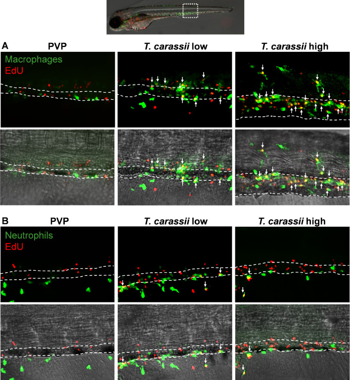
Figure 4—figure supplement 1. Zebrafish were treated as described in Figure 5. (A) A high number of macrophages can be seen around and inside the caudal vein of infected individuals. Especially in high-infected individuals, the majority of cells within the vessel was EdU+, suggesting that in these larvae, dividing macrophages migrated to the vessels. (B) Neutrophils were never observed within the caudal vein and, independently of whether they divided (EdU+) or not, were mostly observed outside or lining the vessel.
Acknowledgments
This image is the copyrighted work of the attributed author or publisher, and
ZFIN has permission only to display this image to its users.
Additional permissions should be obtained from the applicable author or publisher of the image.
Full text @ Elife