IMAGE

Figure 2

ID
ZDB-IMAGE-210625-9
Source
Figures for Leyden et al., 2021
Image
Figure Caption

Figure 2

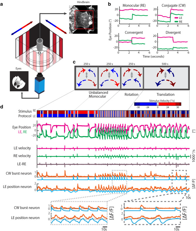
Measurement of cellular calcium signals during quick eye movements. (a) Schematic of the experimental setup. Visually evoked eye movements (image of the eyes in the lower left) and cellular GCaMP calcium signals (optical slice of the hindbrain in the upper right) were recorded in agarose-embedded zebrafish larvae. A black foil in front of the area of binocular overlap enabled independent stimulation of the two eyes with differing stimulus speeds. (b) Example of various horizontal saccade types: monocular, clockwise (CW), convergent (CONV) and divergent (DIV) saccades. The left eye (LE) is shown in magenta, and the right eye (RE) in green. Positive eye positions refer to rightward eye positions. (c) The stimulus protocol consisted of two unbalanced monocular phases, a rotation phase, and finally a translation phase. For each phase, the presented stimulus velocities and directions for the two eyes are illustrated by the size and color of the arrows. During unbalanced stimulation, the absolute stimulus speed was different for the left and the right eyes. For rotation and translation, the absolute speed was the same for the two eyes. (d) Example eye positions and saccadic velocities during stimulation are shown in the top four rows. Only saccadic velocities were of interested for this study and therefore slow-phase velocities were not analyzed here (see “Methods”). Note that we paused stimulus motion after each detected saccade (grey shaded area in the stimulus protocol trace) to decorrelate oculomotor position and velocity signals (see “Methods”). Due to the differential stimulation of the left and the right eye, the eye movements were partially unyoked during unbalanced monocular and translation stimulation (see eye velocity difference trace, LE-RE). The calcium activity (orange) of two example neurons is shown in the lower two traces. One neuron is active during CW saccades (CW velocity regressor in light blue), and the other neuron is coding for rightward eye positions (eye position regressor in light blue). The magnified insets at the bottom allow an easier comparison of activity onset relative to eye position change. Though position neurons can be active at saccadic timepoints (e.g. in the right inset), our saccade-related neurons consistently show their highest calcium fluorescence directly after the saccade.

Acknowledgments
This image is the copyrighted work of the attributed author or publisher, and ZFIN has permission only to display this image to its users. Additional permissions should be obtained from the applicable author or publisher of the image. Full text @ Sci. Rep.