IMAGE

Figure 7

ID
ZDB-IMAGE-210530-15
Source
Figures for Xie et al., 2021
Image
Figure Caption

Figure 7

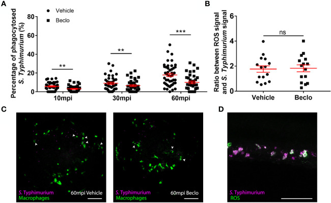
Effect of beclomethasone on phagocytosis of Salmonella Typhimurium and ROS production. At 28 hpf Tg(mpeg1:eGFP) embryos (vehicle- or beclomethasone-treated) were infected with S. Typhimurium through intravenous injection. (A) Percentage of phagocytosed S. Typhimurium wt at 10, 30 and 60 mpi. Confocal microscopy images were taken of the yolk area (as indicated in Figure 3A), and the percentage of bacteria inside macrophages was determined. Statistical analysis, performed by fitting data to a beta-inflated regression with Tukey’s post hoc test, showed that the phagocytic activity of macrophages was significantly inhibited by beclomethasone treatment at 60 mpi, and not at other time points. (B) Ratio between ROS signal and S. Typhimurium signal. Embryos were injected with a ROS biosensor strain. At 4 hpi, confocal microscopy images were taken of the CHT region and the ratio between the ROS-induced fluorescent signal and the signal from a constitutively expressed fluorescent protein in the bacteria was determined. Statistical analysis was performed by two-tailed t-test, indicating no significant difference between the two vehicle- and beclomethasone-treated groups. In panel (A, B), each data point represents a single embryo and the means ± s.e.m. of data accumulated from three independent experiments are shown in red. Statistical significance is indicated by: ns, non-significant; **P<0.01; ***P<0.001. (C) Representative confocal microscopy images of S. Typhimurium wt infected vehicle- and beclomethasone-treated individuals at 60 mpi. Bacteria are shown in magenta, macrophages in green. Arrowheads indicate bacteria phagocytosed by macrophages. Scale bar = 100 μm. (D) Representative confocal microscopy image of a vehicle-treated embryo infected with the S. Typhimurium ROS biosensor strain, showing the bacterial signal (magenta) and the ROS biosensor signal (green). Scale bar = 100 μm.

Acknowledgments
This image is the copyrighted work of the attributed author or publisher, and ZFIN has permission only to display this image to its users. Additional permissions should be obtained from the applicable author or publisher of the image. Full text @ Front Immunol