
|
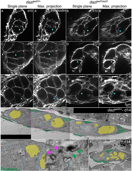
Fig. 8 Single confocal plane of representative dazlae57/+ or dazlae57/ae57 10-14-day gonads labeled with fluorescently conjugated phalloidin (actin) depicting the actin ring. Each stage is represented with a sagittal view (XY) or maximum projection (ten planes of a z-stack). (A,A′,C,C′,E,E′) Actin rings are indicated by a blue arrow in dazlae57/+ 8-14-day gonads. Number of gonads analyzed: 10 days, n=3 (A,A′); 12 days, n=3 (C,C′); 14 days, n=3 (E,E′). (B,B′,D, D′,F,F′) Actin rings at 10 days in dazlae57/ae57 cells. At 12- and 14-days, actin rings are not maintained (blue arrows). Gonads analyzed: 10 days, n=4 (B,B′); 12 days, n=2 (D,D′); 14 days, n=3 (F,F′). (G) TEM image of day 14 wild-type gonad stitched from tiled images (magnification 700×). (H) Magnified image of (G, pink dashed line box) showing a ring canal connecting sister GCs within a cyst. (I) Higher magnification of H (pink dashed line box). (J) TEM image of 14-day dazl mutant gonad stitched from tiled images (magnification 700×). No cysts or ring canals were detected. Pink arrows indicate the centrioles and green arrows indicate the ring canal. Blue arrowheads indicate the germ cell membranes.