
|
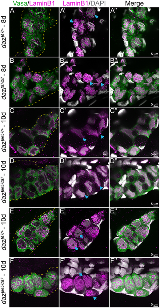
Fig. 5 Single confocal plane of representative dazlΔ7/+ or dazlΔ7/Δ7 day 8 gonads and dazlae57/+, dazlΔ7/+ or dazlΔ7/ae57 at 10 days labeled with Vasa (green), LaminB1 (magenta; nuclear envelope,) and DAPI (gray; DNA). (A-A″) Vasa+ GC of dazlΔ7/+ (n=2) at day 8, with a folded raisin-like nuclear envelope. (B-B″) Vasa+ GC of dazlΔ7/Δ7 (n=3) at day 8, with an unfolded smooth nuclear envelope of Vasa+ GC of dazlae57/+ (C-C″, n=2) at 10 days. (D-D″) The change in nuclear appearance corresponds to amplification during cyst formation and is intact in dazlae57/Δ7. (E-E″) After the transition at 10 days, the nuclei of Vasa+ GC of dazlΔ7/+ remain smooth (n=2). The postmitotic GC nuclei also have a smooth nuclear membrane and a large nucleolus. (F-F″) Vasa+ GC of dazlae57/Δ7 (n=5) at 10 days. Mutant GCs are individuals with a folded nuclear membrane rather than a smooth morphology at 10 days. Yellow dotted line delineates the gonad. Blue arrows indicate each note for the corresponding panel.