
IMAGE
Fig. 3
- ID
- ZDB-IMAGE-210518-3
- Publication
- Paasila et al., 2021 - Ground state depletion microscopy as a tool for studying microglia-synapse interactions
- All Figures
- Figures for Paasila et al., 2021
Image
|
Figure Caption
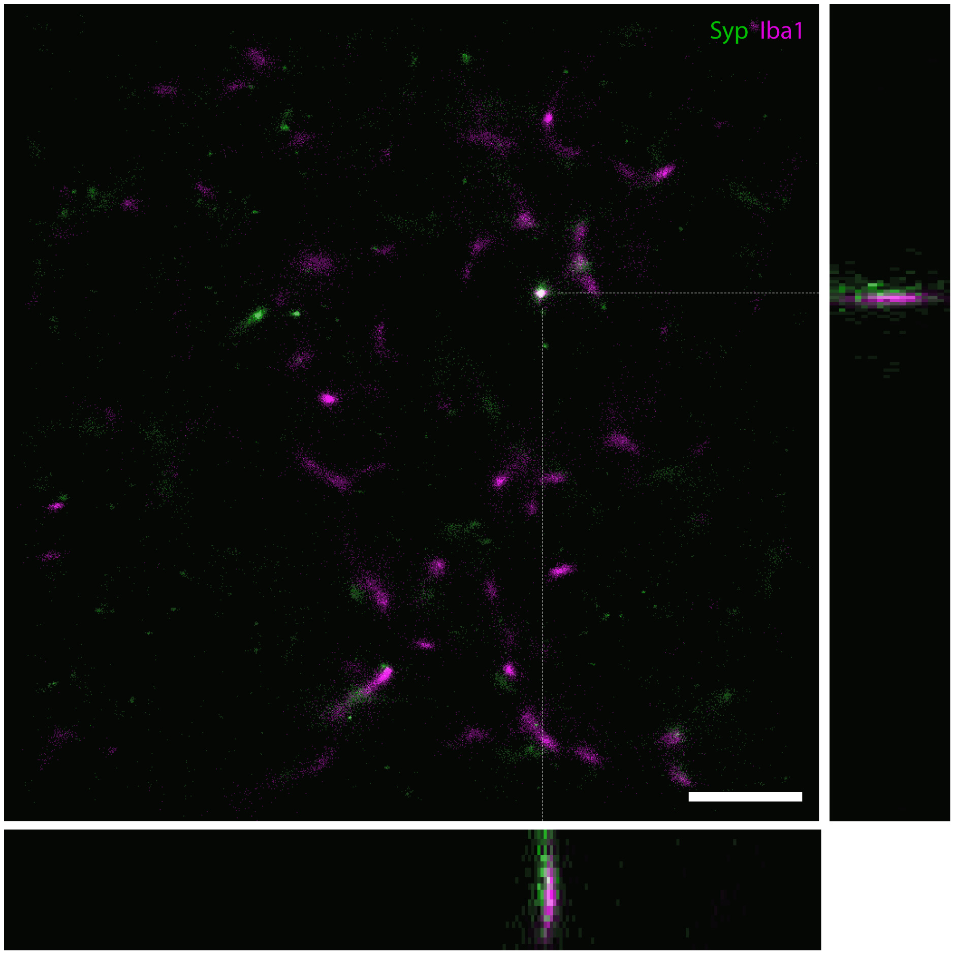
Fig. 3 A 3D GSDIM reconstruction of part of a microglial cell from case AD 2 with a z‐depth of 0.77 µm shown as a maximum intensity projection, with orthogonal views of the XZ and YZ dimensions on the bottom and left of the image, respectively. The orthogonal views have been magnified by 1.99× and cropped for better visualization (each view represents a space that is 0.77 µm in the Z‐direction × 5.17 µm in the X‐ and Y‐direction, respectively). Scale bar = 2.5 µm (XY image only)
Acknowledgments
This image is the copyrighted work of the attributed author or publisher, and
ZFIN has permission only to display this image to its users.
Additional permissions should be obtained from the applicable author or publisher of the image.
Full text @ J. Neurosci. Res.