IMAGE

Figure 8

ID
ZDB-IMAGE-210512-59
Genes
Source
Figures for Sofou et al., 2021
Image
Figure Caption

Figure 8 LysoTracker and GFP‐Lc3 signal are enhanced in <italic>vps16</italic> embryos, suggesting glial dysfunction

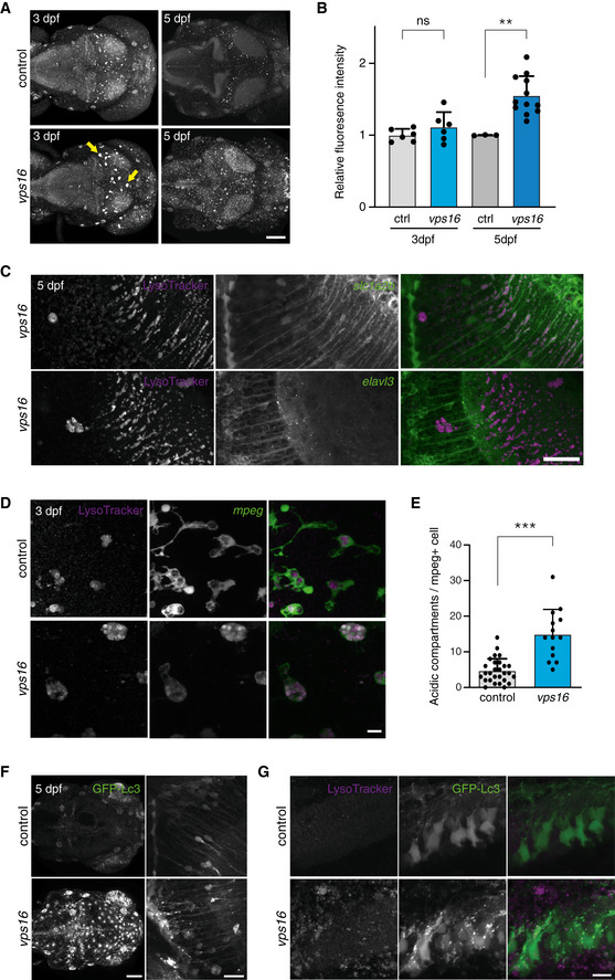
LysoTracker‐stained acidic compartments in zebrafish larvae brains at 3 and 5 dpf. Total signal was increased across the brains of vps16 crispants at 5 dpf, and abnormal, large puncta observed in the optic tectum from 3 dpf (arrows).

LysoTracker signal colocalizes with the astroglial reporter slc1a2b:citrine and does not appear to overlap with the pan‐neuronal reporter elavl3:GCaMP5G.

The puncta indicated in A exist within cells expressing the mpeg1:EGFP transgene, revealing microglia with a grossly altered cellular morphology.

Microglia at 3 dpf contain an increased number of acidic compartments as compared with controls.

Global GFP‐Lc3 signal is increased in unstimulated vps16 embryos (left panel), with clusters of puncta present particularly along the midline of the optic tectum (right panel, magnified view) and anterior hindbrain.

Magnified view of the anterior hindbrain showing numerous GFP‐Lc3 puncta in the vps16 embryo that do not colocalize with LysoTracker.

Data information: Scale bars 100 μm in (A, F left), 30 μm in (C), 20 μm in (F right), and 10 μm in (D, G). Bar graphs represent data as mean ±SEM. Statistical comparisons by Mann–Whitney U‐tests. **P < 0.01; ***P < 0.001. n = 6 each for 3dpf, 3 (ctrl) and 12 (vps16) for 5 dpf in (B). n = 28 (ctrl) and 14 (vps16) in (E). All confocal images representative of a minimum of 6 embryos each.

Figure Data
Acknowledgments
This image is the copyrighted work of the attributed author or publisher, and ZFIN has permission only to display this image to its users. Additional permissions should be obtained from the applicable author or publisher of the image. Full text @ EMBO Mol. Med.