
|
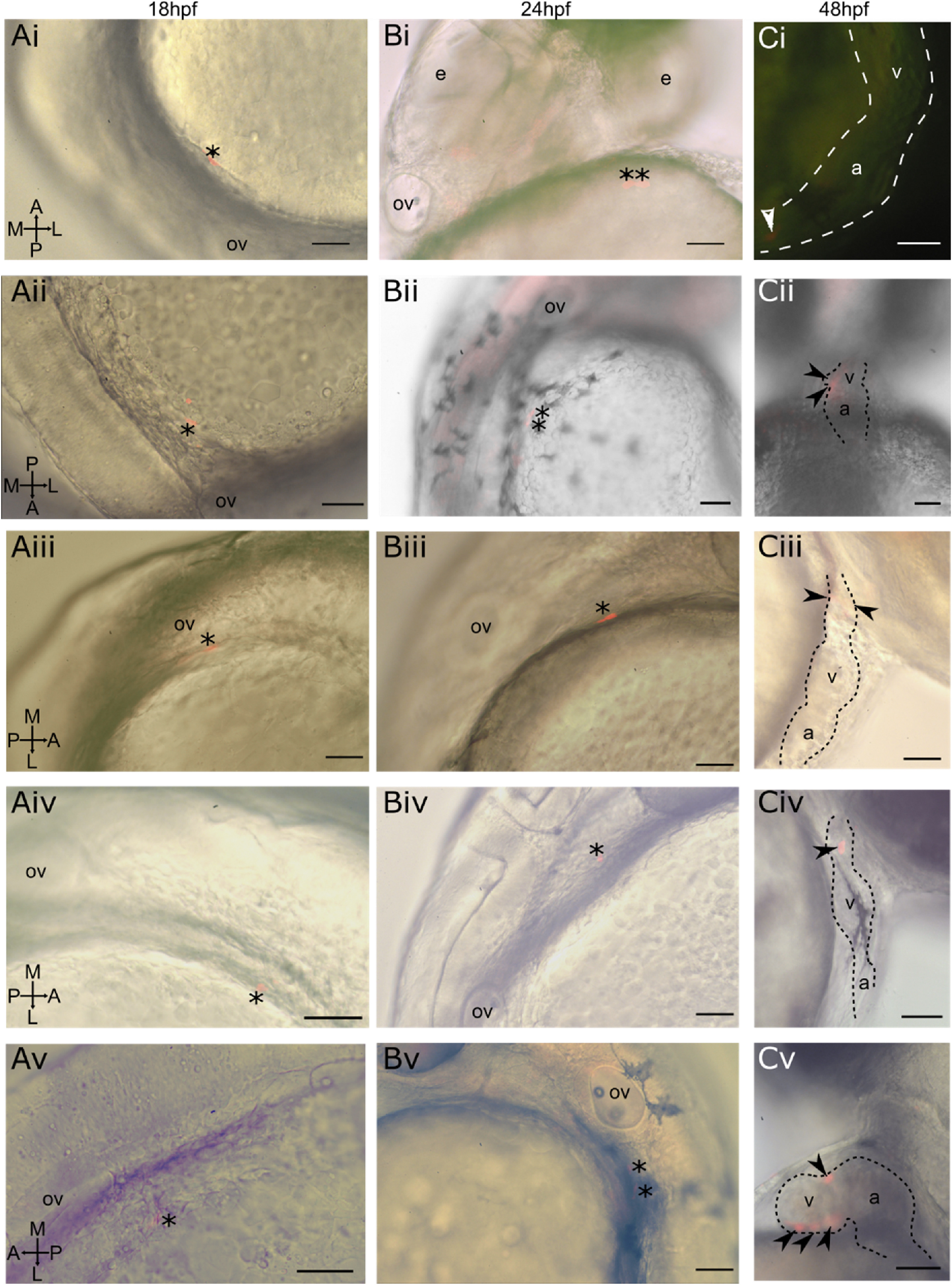
Fig. 3 Photoconverted lateral aLPM cardiac precursor clones do not contribute to the primary heart tube at 24 hpf. i-v Displaying five of 39 examples of wildtype embryos. A- Dorso-lateral view of 18 hpf embryo. Asterisk marks a single cell photoconverted to express red fluorescent Kaede. B- Lateral view of same embryo at 24 hpf. Asterisk marks photoconverted cardiac precursor still within the lateral ALPM and outside of the primary heart tube, currently out of view on the left side of the embryo. C- Lateral view of same embryo at 48 hpf. Arrowheads mark photoconverted cardiac precursors within the larval heart. Ci- Located in in-flow tract; Cii-located in ventricle and atrioventrical canal; Ciii-located in outflow tract; Civ-located in outflow tract; Cv-located in ventricle. a-atrium; v-ventricle; ov-otic vesicle, e−eye. Scale bar 100 μm.
Reprinted from Developmental Biology, 472, Mao, L.M.F., Boyle Anderson, E.A.T., Ho, R.K., Anterior lateral plate mesoderm gives rise to multiple tissues and requires tbx5a function in left-right asymmetry, migration dynamics, and cell specification of late-addition cardiac cells, 52-66, Copyright (2021) with permission from Elsevier. Full text @ Dev. Biol.