
|
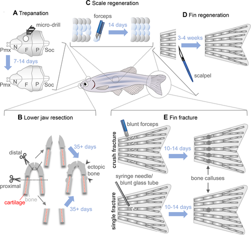
Fig. 5 Models of bone repair and regeneration in adult zebrafish. (A) During skull trepanation, a microdrill is used to destroy bone from the os frontale, which regenerates within 7 to 14 days. (B) Two models of lower jaw resection can be performed: proximal, where both cartilage and bone are removed posterior to the synovial joint using surgical scissors (dotted line), and distal, where only the most anterior part of the bone is removed. Regeneration post resection takes upwards of 35 days and leads to ectopic bone formation in the regenerate. (C) Scale plucking with forceps is a simple method for studying bone regeneration in vivo. Scales can be cultured ex vivo and bone regeneration easily studied dynamically in vivo due to the superficial nature of the injury. (D) Due to its accessibility and the presence of lepidotrichia (bony fin rays), the caudal fin is an excellent model for studying bone regeneration and repair. Epimorphic fin regeneration after amputation requires 3 to 4 weeks until completion. (E) Fin ray fractures are bridged in 10 to 14 days and result in a bone callus, which is then remodeled. Two fin ray fracture models are available: in the crush‐fracture model (top), forceps are used to introduce fractures along the entire width of the fin. In the single‐fracture model, either a syringe needle or blunt glass capillary tube is used to press on an individual bone segment, introducing a single fracture in one or both hemirays. F = os frontale; N = os nasale; P = os parietale; Pmx = os praemaxillare; Soc = os supraoccipitale.