
|
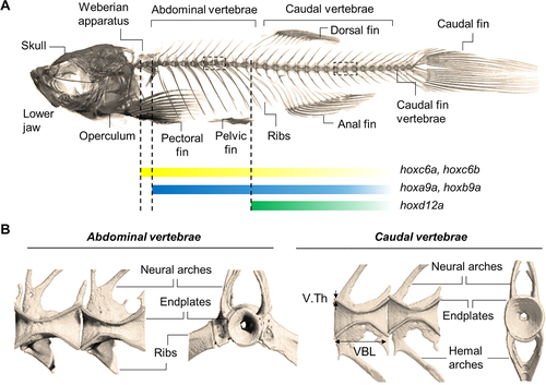
Fig. 1 The zebrafish skeleton. (A) μCT image of the craniofacial and axial skeleton, including the vertebral column composed of the Weberian apparatus, abdominal (also referred to as precaudal or thoracic), caudal, and caudal fin vertebrae. Hox gene expression patterns are indicated. Similar to the mammalian skeleton, ribs in zebrafish are articulated to the abdominal vertebrae and protect the inner organs. (B) Close‐up view of abdominal and caudal vertebrae. Sagittal views of two adjacent double‐cone shaped vertebrae (left) display the neural arches extending dorsally and encompassing the neural canal. Extending ventrally from the abdominal vertebrae ribs are articulated, while the caudal vertebrae extend to hemal arches which encompass the caudal artery and vein. In frontal view (right), the unmineralized vertebral center is revealed which contains notochord and vacuolated soft tissue (not displayed). The ring‐shaped vertebral endplate regions are connected by an IVL (not shown) and correspond to the vertebral growth zone in zebrafish. Parameters, including the VBL, V.Th, and BV/TV, provide valuable measures to quantify the vertebral morphology and structure. BV/TV = bone volumetric fraction; IVL = intervertebral ligament; VBL = vertebral body length; V.Th = vertebral thickness.