
|
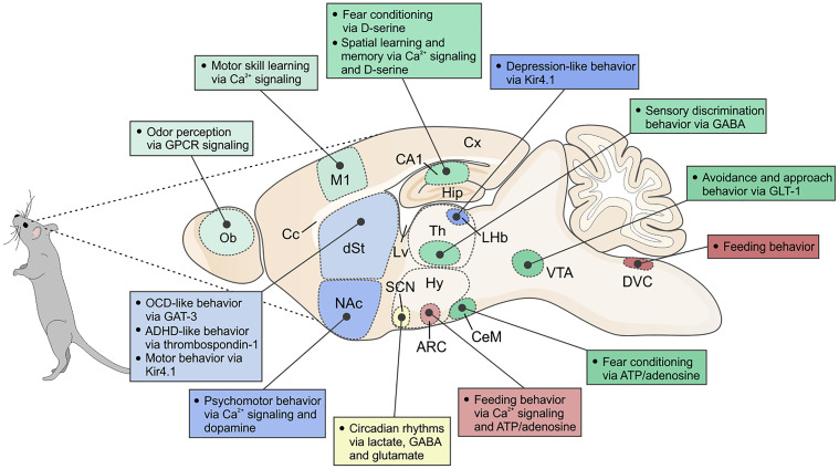
Fig. 3 Schematic of a sagittal section of a mouse brain, depicting various regions and nuclei as well as associated behaviors that have been shown to be regulated by acute astrocytic mechanisms. Ob, olfactory bulb; Cx, cerebral cortex; M1, primary motor cortex; Lv, lateral ventricle; Cc, corpus callosum; dSt, dorsal striatum; NAc, nucleus accumbens; Hip, hippocampus; Th, thalamus; LHb, lateral habenula; Hy, hypothalamus; ARC, arcuate nucleus; SCN, suprachiasmatic nucleus; CeM, central amygdala; VTA, ventral tegmental area; DVC, dorsal vagal complex. In the text, we also consider sleep, but this is not illustrated here because it involves multiple brain nuclei. Furthermore, the cartoon does not include studies where behavioral alterations result over longer periods, such as following deletion of a critical gene within astrocytes or during development and aging.