
Image
|
Figure Caption
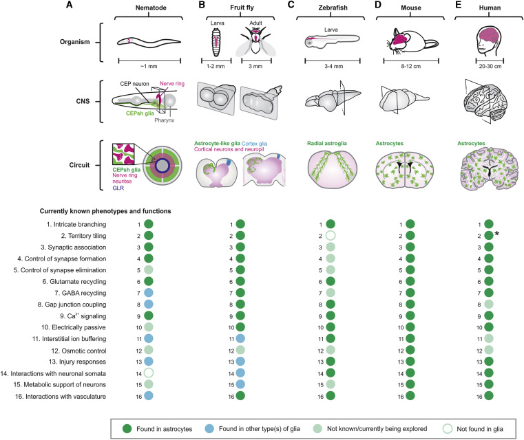
Fig. 1 (A–E) The schematics illustrate the locations of CNS (gray), neuropil (purple), and astrocytes (green) in nematode (A), fruit fly (B), zebrafish (C), mouse (D), and human (E) at the level of organism, CNS, and circuit. Dot plots summarize that 16 well-defined cellular phenotypes/functions of astrocytes are found in astrocytes (green) or other type(s) of glia (blue), not known or currently being explored (light green), or not found in glia (white) in the relevant organism indicated. Some human astrocytes project long unbranched processes that cross cortical laminae (asterisk).
Acknowledgments
This image is the copyrighted work of the attributed author or publisher, and
ZFIN has permission only to display this image to its users.
Additional permissions should be obtained from the applicable author or publisher of the image.
Full text @ Neuron