
Image
|
Figure Caption
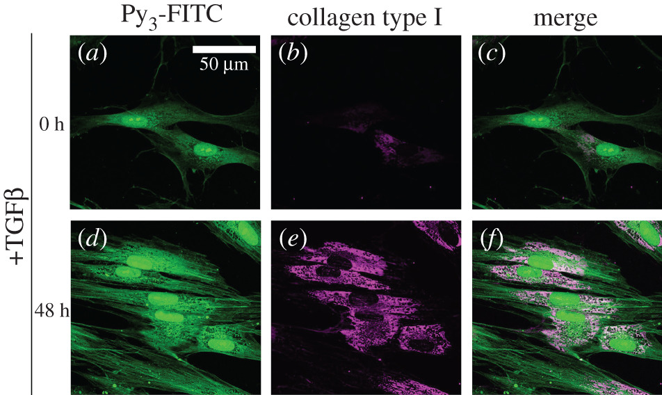
Fig. 6 Py3-FITC can be used to detect TGF-β1-induced collagen type I. HFL-1 cells were treated with TGF-β1 for 0 h (a–c) or 48 h (d–f) and then stained with Py3-FITC (a,d) and collagen type I antibody (b,e), and their merged images are shown (c,f). Collagen type I expression was increased by 48 h of TGF-β1 treatment. The cytoplasmic signal of Py3-FITC was also increased by TGF-β1 and was partially merged with that of collagen type I (c,f). Scale bar in a, 50 μm.
Acknowledgments
This image is the copyrighted work of the attributed author or publisher, and
ZFIN has permission only to display this image to its users.
Additional permissions should be obtained from the applicable author or publisher of the image.
Full text @ Open Biol.