
|
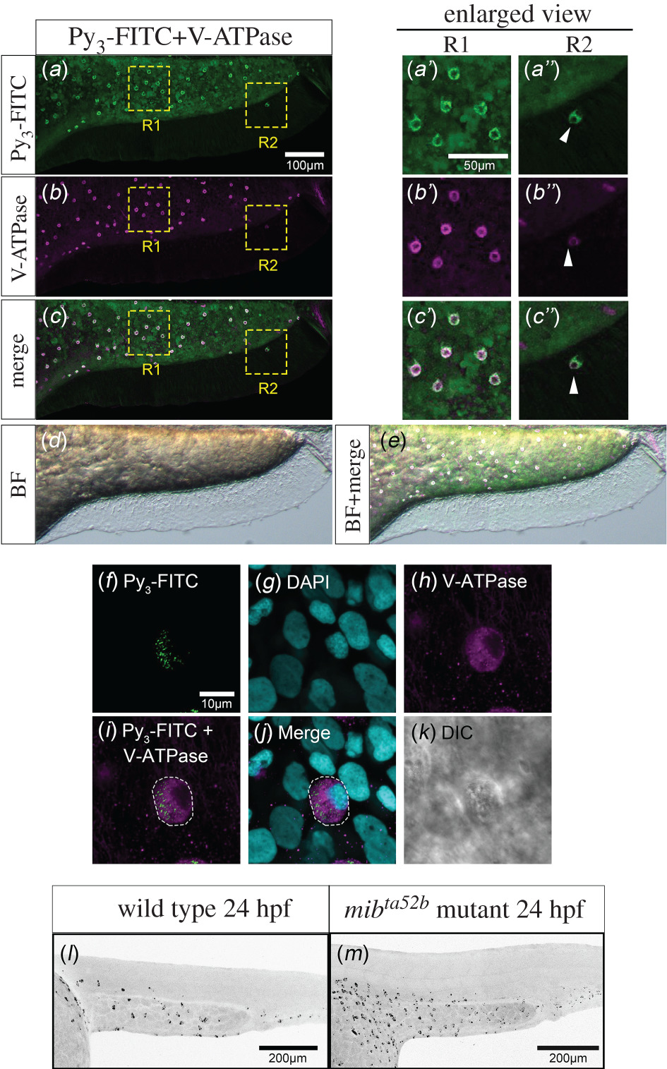
Fig. 2 Py3-FITC-positive cells on the yolk and trunk surface are HR cells. Lateral views of Py3-FITC and anti-V-ATPase antibody double-stained zebrafish embryos at 3 dpf. (a) Py3-FITC-stained cells and (b) V-ATPase-expressing cells mostly overlapped (c). (d) Bright field image. (e) The overlaid image of c and d. Enlarged views of R1 and R2 region of Py3-FTIC staining (a′, a″), V-ATPase staining (b′, b″) and merged view (c′, c″) are also shown here. The white arrowhead indicates Py3-FITC-positive HR cell on the ventral fin (a″, b″, c″). (f–k) High magnification view of Py3-FITC-stained HR cells in 3 dpf fixed zebrafish. The projected image of Py3-FITC staining (f), the projected image of DAPI staining (g), the projected image of V-ATPase antibody staining (h), The overlaid image (i) of f and h, merged view (j) and DIC image (k) are shown. (l,m) Lateral views of Py3-FITC-stained wild-type (l) and mibta52b-mutant (m) zebrafish embryos at 24 hpf. Embryos were treated with 10 μM Py3-FITC for 5 h from 19 hpf (l, m). Py3-FITC-positive cells were significantly increased in Notch signalling-deficient mibta52b embryos. Scale bars in a, 100 µm; a′, 50 µm; f, 10 µm; l, m, 200 µm.