
|
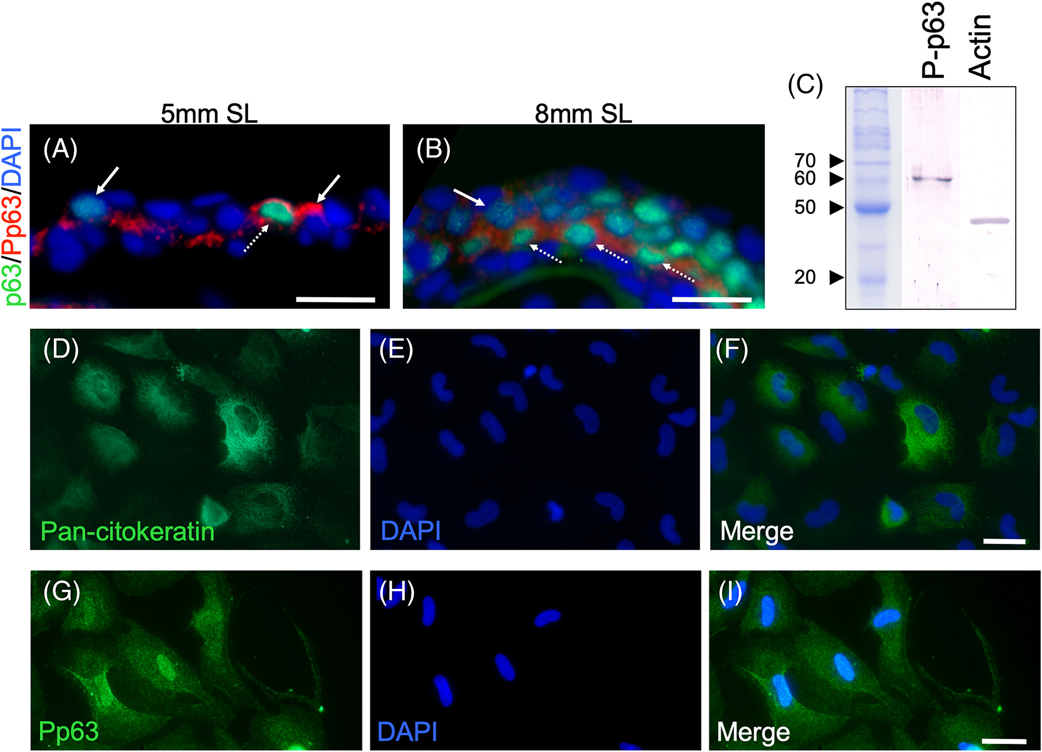
Fig. 5 Presence of phosphorylated p63 during epidermal stratification. A, Double‐labeling of longitudinal sections of 5‐mm‐SL larvae with anti‐p63 and anti‐Pp63. B, Immunostaining with anti‐p63 and anti‐Pp63 at 8 mm SL. A‐B, Dotted arrows point to cells with Pp63 and high levels of p63, while arrows mark cells with Pp63 and low or null levels of p63. C, Western blot of Pp63, and Actin as a loading control. D‐I, Epidermal cells from zebrafish in primary cell culture labeled with anti‐p63 and anti‐Pp63. A and B Images were obtained with a 40X oil objective, while D‐I were obtained with a 63X oil objective. Scale bar in A, B, and D‐I is 20 μm