
|
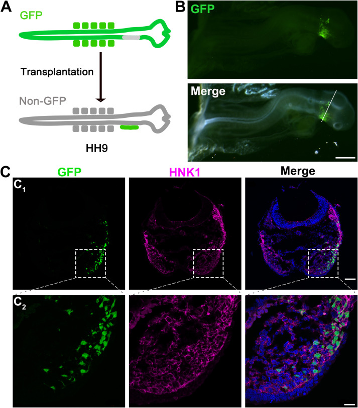
Fig. 3 Fig. 3. Migration of GFP+ NC cells ventrally in GFP− host chicken after the insertion of the GFP+ neural fold. A: A schematic graph illustrating the insertion of GFP+ neural fold to GFP− host chicken embryo. B: Photomicrographs of a GFP+/GFP− chicken chimera at 1 DPS. Top: fluorescent image to show GFP signals; Bottom: merged fluorescent and bright-field images. C: Single-plane laser scanning confocal images of a section (C1) from the position indicated by white line in B and higher power images (C2) from the area indicated by dashed square shown in C1. Sections were immunoreacted for NC cell marker HNK1 (magenta). Scale bars: 500 μm in B; 80 μm in C1 and 20 μm in C2.
Reprinted from Developmental Biology, 471, Yu, W., Wang, Z., Marshall, B., Yoshida, Y., Patel, R., Cui, X., Ball, R., Yin, L., Kawabata, F., Tabata, S., Chen, W., Kelsh, R.N., Lauderdale, J.D., Liu, H.X., Taste buds are not derived from neural crest in mouse, chicken, and zebrafish, 76-88, Copyright (2020) with permission from Elsevier. Full text @ Dev. Biol.