IMAGE

Fig. 3

ID
ZDB-IMAGE-210315-3
Source
Figures for Hilbert et al., 2021
Image
Figure Caption

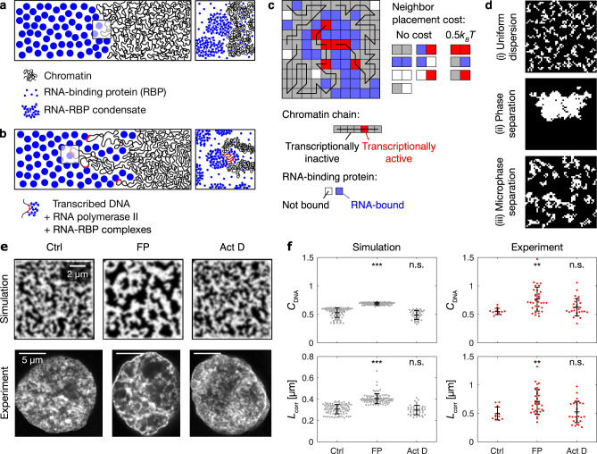
Fig. 3 A microemulsion model reproduces key features of euchromatin organization.

a Model mechanism 1: segregation of RNA-RBP condensates from chromatin. b Model mechanism 2: tethering of RNA-RBP complexes to transcriptionally active chromatin, forming amphiphilic connections. c Illustration of the lattice model for euchromatin organization, indicating chromatin chains and RNA-binding proteins with different states, as well as the costs for placing different types of lattice sites next to each other. See Supplementary Table 1 for model parameters. d Configurations of chromatin (white) in long-term simulations of the lattice model with (i) no RNA and no transcription, (ii) RNA and no transcription, and (iii) RNA and transcription. e Concentration profiles from simulations with example STED nuclear mid-sections from corresponding experimental conditions: control treatment (Ctrl), flavopiridol (FP), actinomycin D (Act D). The same results were obtained in four independent experiments. f Comparison of DNA image contrast (CDNA) and correlation length (Lcorr) from simulations and STED mid-nuclear sections. Mean ± SD, ** indicates P < 0.01, *** indicates P < 0.001, n.s. indicates P ≥ 0.05 (CDNAP values <10−5, 0,33 from a two-sided permutation test, LcorrP values 0.003, 1.09 from a two-sided permutation test, P values with Bonferroni correction for multiple testing and resampling n matched to experiments, n = 96,93,46 simulations, for a description of the statistics of experiments, see Fig. 2c).

Acknowledgments
This image is the copyrighted work of the attributed author or publisher, and ZFIN has permission only to display this image to its users. Additional permissions should be obtained from the applicable author or publisher of the image. Full text @ Nat. Commun.